Summary
Hydrocephalus is an elevation in intracranial pressure (ICP) caused by a disturbance in the flow, absorption, or production of cerebrospinal fluid (CSF), resulting in CSF accumulation and progressive dilation of the cerebral ventricles. In children it most often reflects impaired CSF circulation.
The clinical picture depends on whether the cranial sutures are still open: infants develop macrocephaly with a bulging fontanelle, whereas older children present with features of raised ICP. The mainstay of treatment is surgical CSF diversion with a ventriculoperitoneal (VP) shunt.
Definition
Hydrocephalus is an elevation in intracranial pressure due to a disturbance in the circulation of cerebrospinal fluid (CSF), with accumulation of CSF and dilation of the ventricular system. The underlying disturbance falls into one of three mechanisms:
- Impaired CSF circulation (obstruction of flow) — the most common cause.
- Impaired CSF absorption — at the arachnoid granulations.
- Excessive CSF production.
CSF Physiology & Pathway
In an adult the normal CSF volume is about 150 mL, with roughly 25% within the ventricular system; in an infant the total CSF volume is about 50 mL. Normal CSF production is 400–500 mL daily, so the entire volume turns over several times a day — any mismatch between production and absorption rapidly raises ICP.
CSF follows a fixed one-way circuit: the choroid plexus of the paired lateral ventricles → the two foramina of Monro → third ventricle → aqueduct of Sylvius → fourth ventricle → three exit openings (two lateral foramina of Luschka and the median foramen of Magendie) → the spinal and intracranial subarachnoid space over the cerebral convexities → absorbed into the venous sinuses through the arachnoid granulations. Knowing this circuit is essential: an obstruction at any point produces non-communicating hydrocephalus, dilating every cavity proximal to the block.
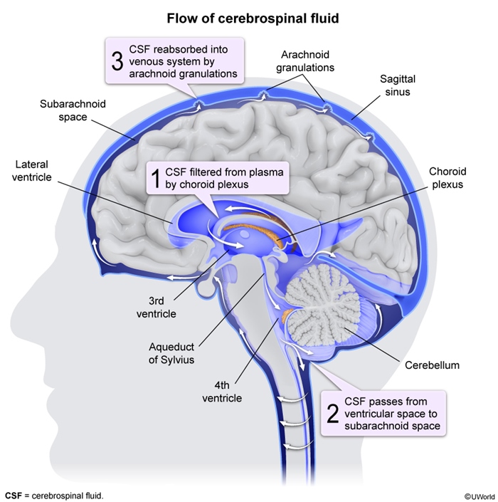
| CSF Flow Pathway – جملة تذكرية | |
“Charles Monroe Threw A Frisbee, Luschka-Magendie Sailed Across”
|
جملة تذكرية |
Types & Classification
Hydrocephalus is classified by CSF dynamics into three patterns. The key distinction for exams is whether the ventricular pathways still communicate — and, if not, where the obstruction lies.
These three categories comprise non-communicating (obstructive) hydrocephalus, communicating (nonobstructive) hydrocephalus, and hydrocephalus ex vacuo, a distinction that fundamentally determines the underlying pathophysiology and appropriate therapeutic approach.
| Classification of Hydrocephalus: By CSF dynamics | |
| 1. Non-communicating | Obstructive |
| Mechanism | Obstruction within the ventricular system; CSF builds up proximal to the block and the ventricular pathways no longer freely communicate |
| Typical causes | Aqueductal stenosis, Chiari II, Dandy–Walker, tumors obstructing the CSF pathway |
| 2. Communicating | Nonobstructive |
| Mechanism | All ventricular pathways communicate normally; the problem is increased CSF production or decreased absorption at the arachnoid granulations |
| Typical causes | Post-hemorrhagic or post-meningitic scarring of granulations; CSF-producing tumors |
| 3. Hydrocephalus ex vacuo | Not true hydrocephalus |
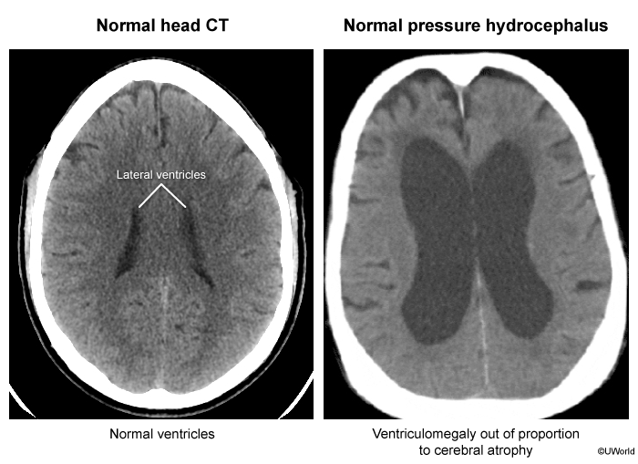
| Mechanism | Ventricular enlargement caused by loss of brain parenchyma (atrophy), NOT a disturbance of CSF dynamics; intracranial pressure is normal |
| Management | Shunting is NOT indicated |
| فخ امتحاني – Exam Trap | |
|
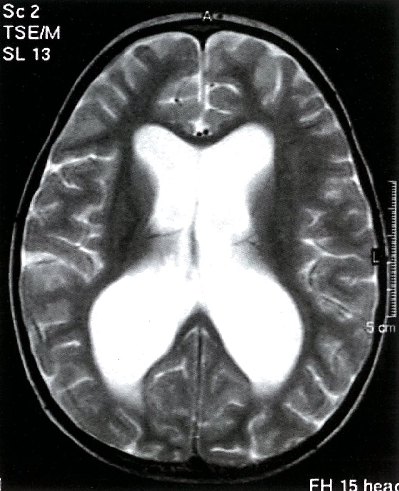
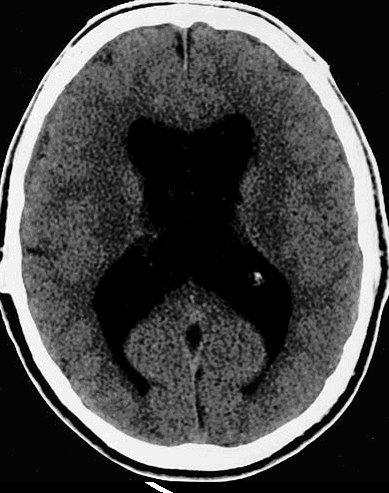
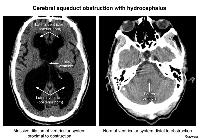
Pay attention to the distinction between communicating and non-communicating hydrocephalus: in non-communicating hydrocephalus, there is an obstruction, so only the ventricles proximal to the blockage enlarge (for example, in aqueductal stenosis, the lateral and 3rd ventricles enlarge while the 4th remains normal). In contrast, in communicating hydrocephalus, all ventricles enlarge because the problem lies with absorption or production, not with flow. |
تذكر |
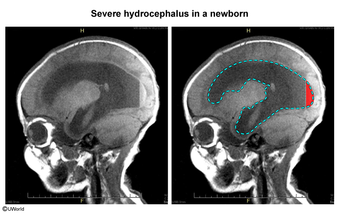
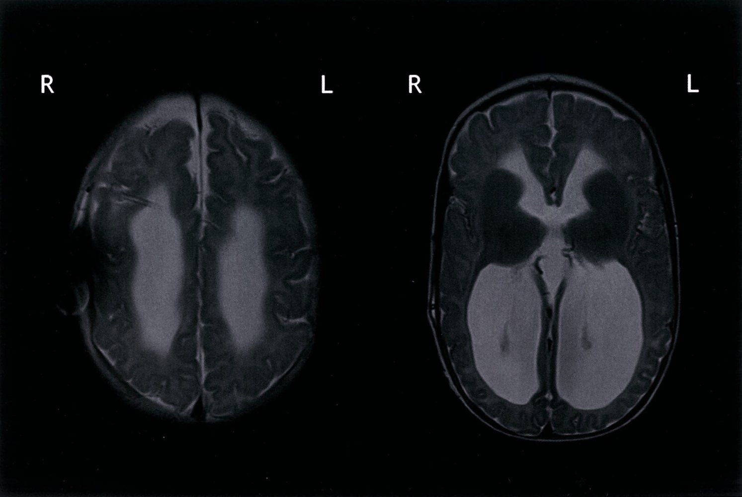
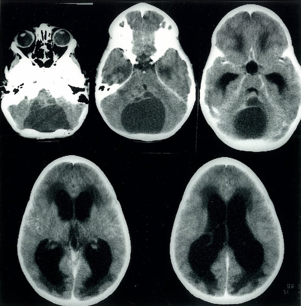
Etiology — Congenital & Acquired Causes
Etiological causes are divided into congenital and acquired groups.
1. Congenital causes
| Congenital Causes of Hydrocephalus | |||
|---|---|---|---|
| Feature | Chiari II malformation | Dandy–Walker malformation | Congenital aqueductal stenosis |
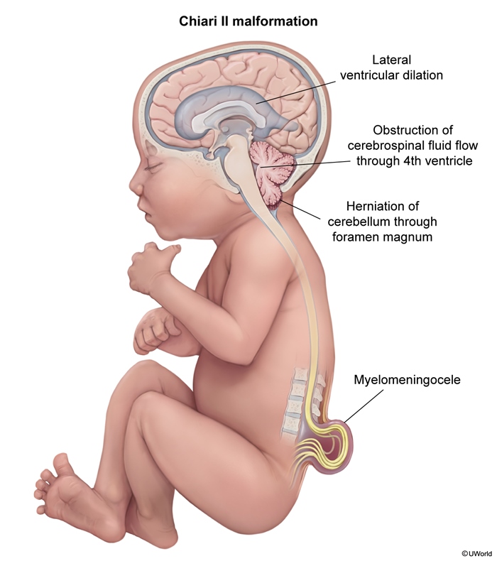
| Core defect | Downward displacement of cerebellum & medulla through the foramen magnum | Absent or hypoplastic cerebellar vermis + cystic enlargement of the 4th ventricle | Narrowing of the aqueduct of Sylvius |
| How it blocks CSF | Herniated hindbrain obstructs CSF flow | Cystic 4th ventricle obstructs CSF outflow | Obstruction between the 3rd and 4th ventricles |
| Key association | Lumbosacral myelomeningocele | Posterior fossa cystic enlargement | X-linked form → thumb abnormalities & other CNS anomalies (e.g., spina bifida) |
| Exam note | — | — | Most common cause of non-communicating hydrocephalus |
Chiari II malformation shows hindbrain herniation through the foramen magnum, while aqueductal stenosis produces a characteristic pattern of proximal ventricular dilation on imaging.
| فخ امتحاني – Exam Trap | |
|
Do not confuse Chiari II with Dandy–Walker: Chiari II involves herniation of the cerebellum and medulla downward through the foramen magnum and always comes with lumbosacral myelomeningocele, whereas Dandy–Walker's problem is in the posterior fossa — the vermis is absent or hypoplastic with a cyst in place of the 4th ventricle. |
تذكر |
2. Acquired causes
The three most commonly tested acquired causes are:
- Intraventricular hemorrhage — most common in preterm infants.
- Bacterial meningitis — inflammatory scarring impairs CSF absorption.
- CSF-producing brain tumors — overproduction of CSF.
See the congenital hydrocephalus causes and pediatric red-flags for how these etiologies map onto the early warning signs of macrocephaly and a bulging fontanelle.
Clinical Features
A head circumference that crosses percentile lines, or exceeds the 97th percentile for age, is the cardinal sign. The remaining findings depend on whether the cranial sutures are still open or already fused.
| Clinical Features by Age Group | ||
|---|---|---|
| Finding | Infants — open sutures & fontanelles | Older children — closed sutures (raised-ICP picture) |
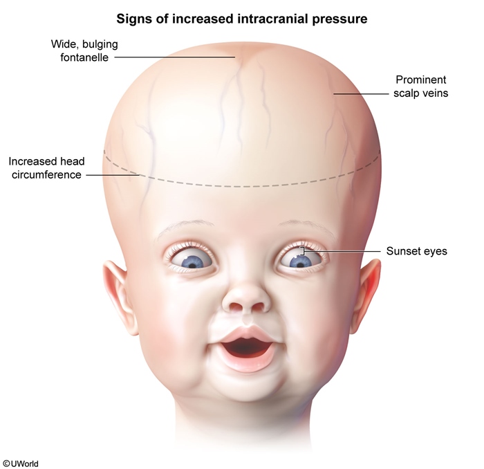
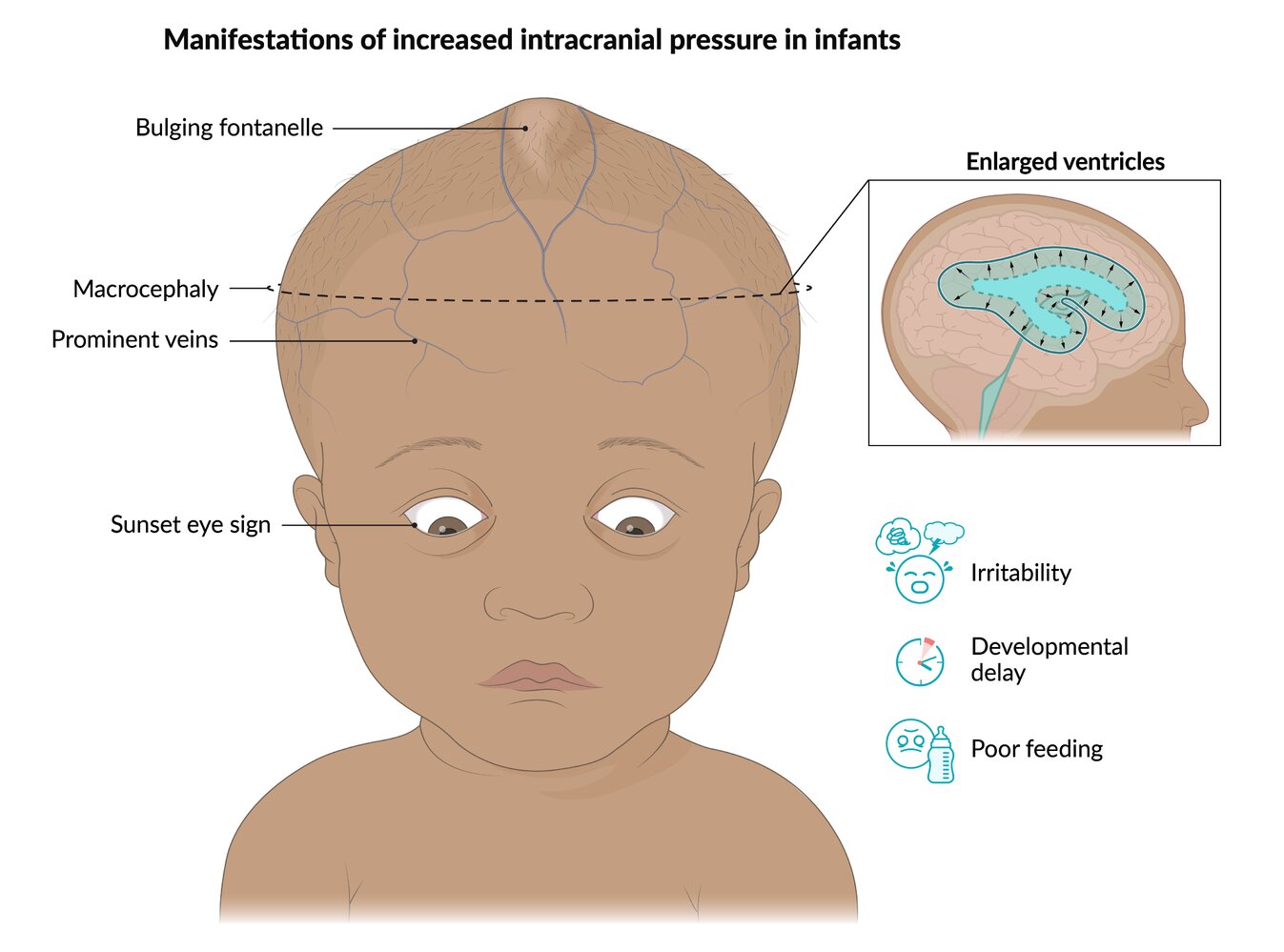
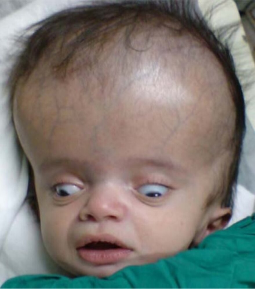
| Head & scalp | Macrocephaly; head circumference crossing percentile lines or >97% for age; rapidly enlarging head; prominent scalp veins | Head size usually normal; features of raised ICP dominate |
| Sutures / fontanelle | Tense, bulging anterior fontanelle; widely spaced or split sutures | Fused sutures — no fontanelle signs |
| Eyes | Sun-setting eyes (tonic downward deviation of both eyes) | Papilledema; unilateral sixth (abducens) nerve palsy |
| Behaviour & GI | Poor feeding, irritability, decreased activity, vomiting, developmental delay | Headache; nausea and vomiting |
| Reflexes / vitals | — | Brisk deep tendon reflexes with a downward plantar response; hypertension with bradycardia (Cushing response) |
| ملاحظة سريرية – Clinical Note | |
|
Sun-setting eyes result from compression of the upward gaze centre in the midbrain by the dilated 3rd ventricle, causing tonic downward deviation of the eyes—a classic sign of hydrocephalus in infants. |
ملاحظة |
The causes and complications of raised intracranial pressure lays out how headache, vomiting, and the Cushing response fit into the broader picture of decompensating ICP.
Evaluation, Management & Prognosis
Evaluation
An increasing head circumference together with signs or symptoms of raised ICP mandates urgent neuroimaging: a head ultrasound through the open fontanelle in infants, or a head CT or MRI in older children.
Management
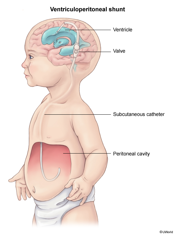
Definitive treatment is surgical CSF diversion with a ventriculoperitoneal (VP) shunt, which drains CSF from a lateral ventricle to the peritoneal cavity. The two principal shunt complications are shunt infection and shunt obstruction. In older children, acetazolamide may be used adjunctively to lower ICP by reducing CSF production.
| فخ امتحاني – Exam Trap | |
|
Hydrocephalus ex vacuo is not true hydrocephalus — the ventricles enlarge because of brain atrophy (loss of parenchyma), not due to elevated pressure. Thus, the ICP remains normal and shunt placement is not indicated. |
تذكر |
Prognosis
Outcome depends on the underlying cause and the success of intervention. Patients with aqueductal stenosis have the best cognitive outcome; those with Chiari II malformation may have low-normal intelligence with language disorders; and patients with X-linked hydrocephalus frequently have cognitive delays.
Key Points for Exams – نقاط مهمة للامتحانات
| Key Points for Exams – نقاط مهمة للامتحانات | |
|
تذكر |
احصل على التجربة الكاملة
اشترك للوصول لفيديوهات الشرح التفصيلي والبطاقات التعليمية التفاعلية وأسئلة الممارسة مع تتبع التقدم.