Summary
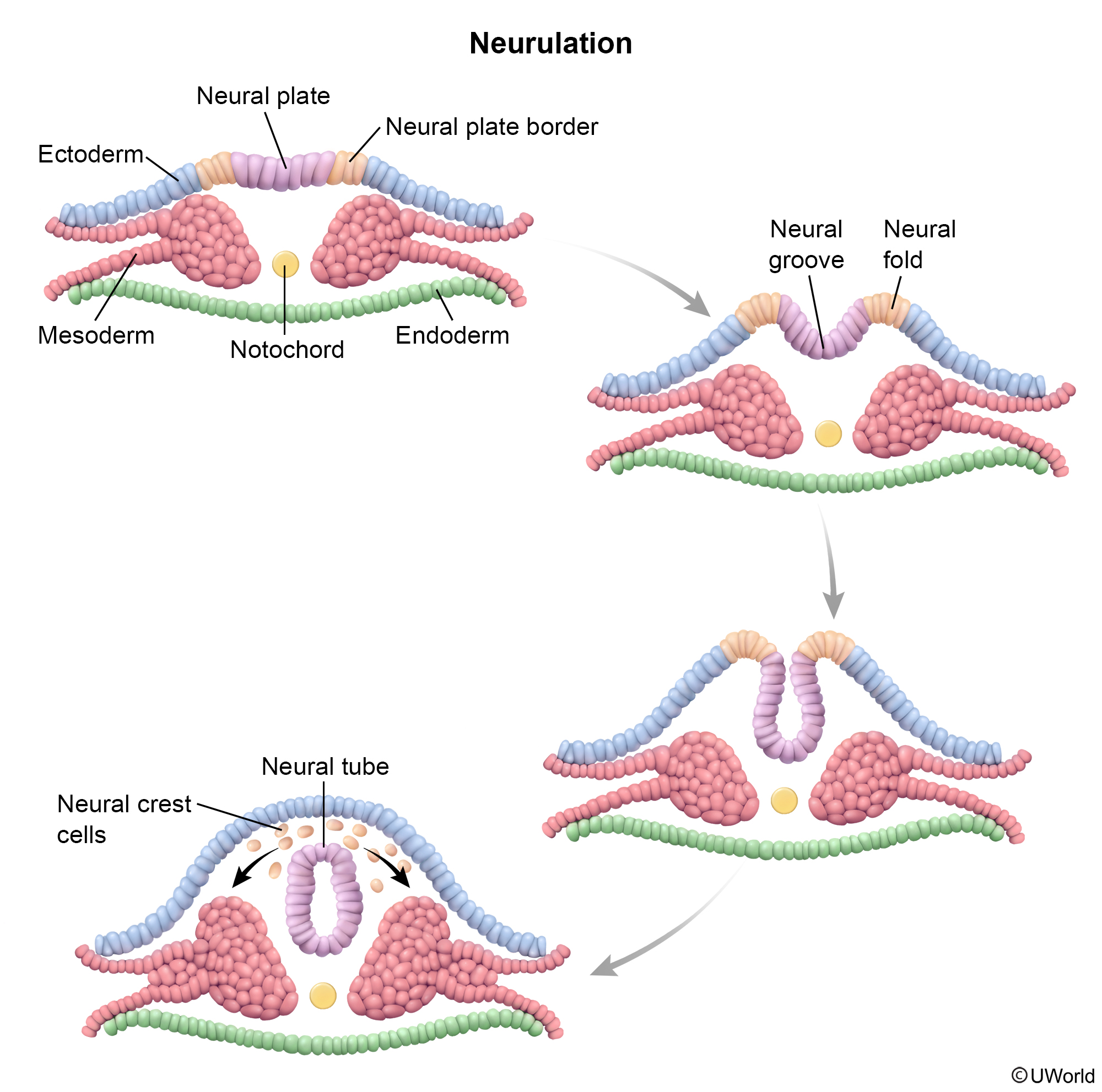
Neural tube defects (NTDs) are among the most common and most heavily tested congenital anomalies of the central nervous system. They arise from incomplete closure of the neural tube during the 3rd–4th week of embryonic development (neurulation). Depending on where closure fails, the resulting defect is classified as cranial — anencephaly (lethal) or encephalocele — or caudal — the spina bifida spectrum (occulta → meningocele → myelomeningocele).
The single most important modifiable risk factor is folic acid deficiency; other contributors include antiepileptic drugs (valproate, carbamazepine), maternal diabetes, obesity, and hyperthermia.
Prenatal screening relies on elevated maternal serum alpha-fetoprotein (MSAFP) in the second trimester followed by confirmatory ultrasound; elevated AFP in amniotic fluid with acetylcholinesterase further supports the diagnosis of open NTDs. Prevention centres on periconceptional folic acid supplementation — 0.4 mg/day for all women of childbearing age and 4 mg/day for those with a prior affected pregnancy.
Overview & Definition
Neural tube defects (NTDs) are a group of congenital malformations caused by failure of neural tube closure during the 3rd–4th week of embryogenesis (neurulation). They are among the most common serious congenital anomalies of the CNS.
Closure proceeds in both directions along the neural tube: the anterior neuropore normally closes by ~day 25 and the posterior neuropore by ~day 27–28. Where closure fails determines the lesion:
- Anterior (cranial) failure → Anencephaly (and encephalocele).
- Posterior (caudal) failure → Spina bifida, including myelomeningocele.
A key organising concept is open vs closed defects: in open defects neural tissue communicates with amniotic fluid, so alpha-fetoprotein (AFP) leaks out and rises; closed (skin-covered) defects keep AFP normal.
Epidemiology, Etiology & Prevention
Epidemiology: NTDs occur in approximately 1 in 1000 live births (0.1%).
Etiology: the cause is multifactorial — genetic, nutritional, environmental and teratogenic factors interact. Periconceptional folate status is central: adequate folic acid supplementation prevents roughly 50% of NTDs, including spina bifida and anencephaly.
Risk factors include a previous NTD-affected pregnancy (strongest single factor), inadequate folic acid intake, and maternal diabetes mellitus. The highest-yield teratogens are valproic acid, carbamazepine, phenytoin, and methotrexate (antiepileptics and folate antagonists). Valproic acid alone raises NTD incidence from the baseline 0.1% to 1–2%.
| Mnemonic – جملة تذكرية: NTD Risk Factors = "FAD" | |
➕ Add a prior NTD-affected pregnancy — the single strongest individual risk factor. |
جملة تذكرية |
Prevention: folic acid must begin before conception and continue through the first 12 weeks of gestation. Dosing is risk-stratified:
| Daily folic acid dose | Indication / timing | |
| Average risk | 0.4 mg (400 µg) PO daily | All women planning pregnancy; start before conception, continue through the first 12 weeks of gestation |
| High risk | 4 mg PO daily | Previous NTD-affected pregnancy, maternal diabetes, or antiepileptic (e.g., valproate) therapy |
Refer to the NTD risk-factor, prenatal screening and prevention overview for a consolidated summary of culprits, screening modalities and supplementation guidance.
Spina Bifida Subtypes & Complications
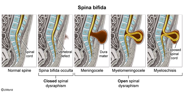
Spina bifida is any failure of bony fusion in the posterior midline of the vertebral column. Three subtypes are recognised in order of increasing severity: spina bifida occulta, meningocele, and myelomeningocele.
1. Spina bifida occulta
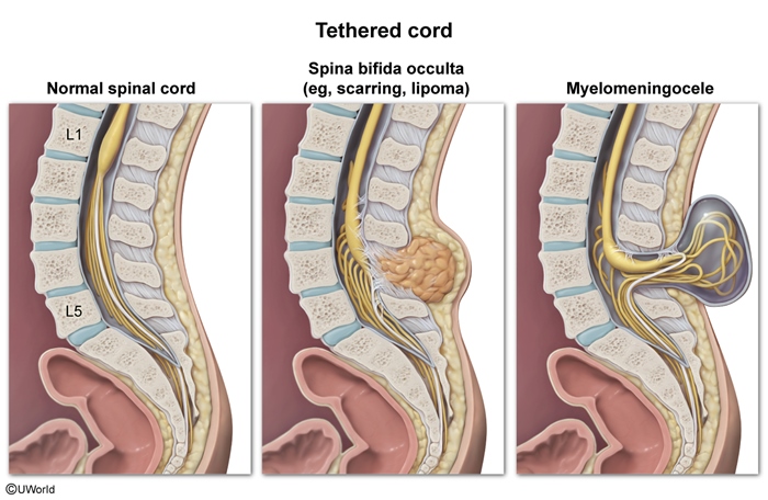
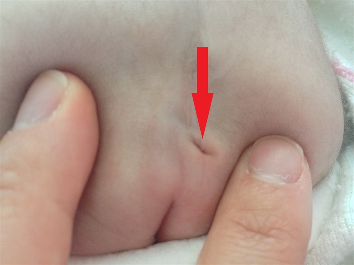
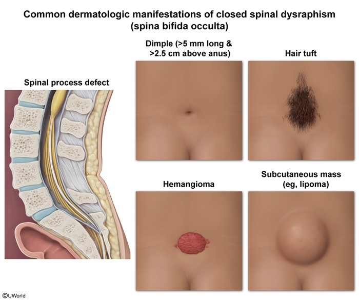
Spina bifida occulta is the most common subtype. There is incomplete vertebral fusion but no herniation of neural tissue. The defect is skin-covered and is often marked externally by a hairy patch, lipoma or dimple over L5–S1. A neurologic deficit is usually absent, although some patients have constipation, leg weakness or a neurogenic bladder. Because it is a closed defect, maternal serum AFP is normal. Associations include a dermoid sinus, tethered cord and other cord anomalies. No treatment is required.
2. Meningocele
Meningocele is the rarest subtype. Only the meninges herniate through the bony cleft, producing a CSF-filled sac that is usually skin-covered and transilluminates. The neurologic deficit is absent or mild, and maternal serum AFP is normal when the lesion is skin-covered. The anterior sacral form can present with constipation or bladder dysfunction. Treatment is surgical repair.
3. Myelomeningocele
Myelomeningocele is the most severe and clinically most important subtype. Both the meninges and the spinal cord / nerve roots herniate through the cleft, frequently as an exposed neural placode (an open defect). A neurologic deficit is present distal to the lesion — the lower the lesion, the milder the deficit. Maternal serum AFP is elevated because the defect is open. It is associated with Chiari II malformation plus hydrocephalus (~90%), hydrosyringomyelia, and orthopedic and genitourinary deficits. Management is urgent surgical repair within 24 hours of birth (or in utero).
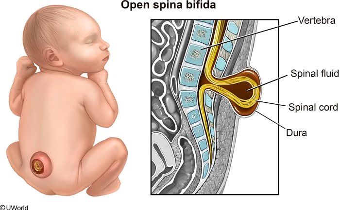
- Most severe and clinically most important NTD: failure of neural tube closure → protrusion of meninges, CSF and spinal cord through a membranous sac, usually in the lumbosacral region; strongly linked to maternal folate deficiency.
- A fluctuant midline mass that involves the nerve roots, spinal cord, meninges, vertebral bodies and skin.
- Neurologic deficits distal to the lesion: complete paraplegia with lesions above L3, versus preserved ambulation and variable bowel/bladder incontinence at S3 and below. Rule: the lower the lesion, the milder the deficit.
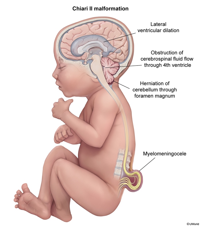
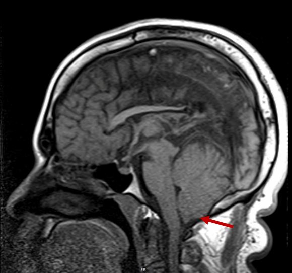
- Chiari II malformation with obstructive hydrocephalus in ~90% of lumbosacral lesions — inferior displacement of the medulla and cerebellum through the foramen magnum.
- Other associations:
- Cervical hydrosyringomyelia
- Neuronal migration defects (gyral anomalies, agenesis of the corpus callosum)
- Orthopedic deformities and insensate fractures.
- Genitourinary neurologic deficits.
| Important – فخ امتحاني | |
|
Always associate Myelomeningocele with Chiari II malformation and hydrocephalus, because it alters management and the patient may require shunting. |
تذكر |
| Spina Bifida: Subtypes Comparison | |||
|---|---|---|---|
| Feature | Spina bifida occulta | Meningocele | Myelomeningocele |
| Frequency | Most common | Rarest | Most severe and clinically most important |
| Herniation | No herniation of neural tissue | Only meninges herniate | Meninges and spinal cord / nerve roots herniate |
| Defect type | Closed defect | Usually skin-covered | Open defect |
| Skin coverage | Skin-covered; marked by hairy patch, lipoma or dimple over L5–S1 | CSF-filled sac, usually skin-covered, transilluminates | Exposed neural placode; fluctuant midline mass involving nerve roots, spinal cord, meninges, vertebral bodies and skin |
| Neurologic deficit | Usually absent; some patients have constipation, leg weakness or neurogenic bladder | Absent or mild | Present distal to lesion; lower lesion = milder deficit (complete paraplegia above L3; preserved ambulation at S3 and below) |
| Maternal serum AFP | Normal | Normal when skin-covered | Elevated |
| Associations & Complications | Dermoid sinus, tethered cord, other cord anomalies | Anterior sacral form: constipation or bladder dysfunction | Chiari II malformation with obstructive hydrocephalus (~90%), hydrosyringomyelia, neuronal migration defects, orthopedic deformities, genitourinary deficits |
| Management | No treatment required | Surgical repair | Urgent surgical repair within 24 hours of birth (or in utero) |
Encephalocele
An encephalocele is herniation of the brain, its coverings, or both, through a skull defect. It is occipital in ~75% of cases, with males and females affected equally.
- The herniated sac may contain meninges, CSF, or dysplastic neural tissue.
- Sac size does not correlate with the amount of brain tissue present — transillumination gives a better bedside assessment.
- If the sac contains meninges only, development is usually normal with no CNS deficits; significant brain tissue involvement predicts poor neurologic outcome.
- MRI defines how much brain tissue is within the encephalocele and guides prognosis.
Diagnosis: prenatal ultrasound and/or an elevated maternal AFP; note that AFP may be normal if the defect is covered by skin/scalp (closed lesion). Treatment is prompt surgical removal of the sac with closure of the defect.
Anencephaly
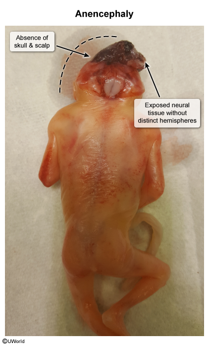
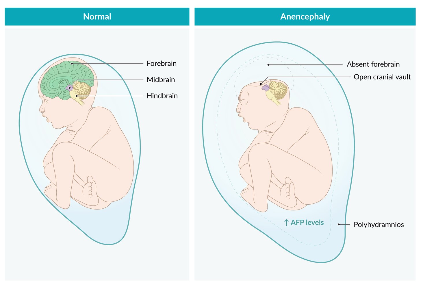
Anencephaly occurs when the anterior neuropore fails to close (normally complete by day 24 post-conception). The result is absence of both cerebral hemispheres, usually with no hypothalamus; the resulting hypoplastic pituitary gland causes failure of end-organ development (e.g., adrenal insufficiency).
- Large parts of the cranium are missing — typically the occipital bones, sometimes the frontal and parietal bones.
- Associated facial and ocular malformations are common; girls are affected more often than boys.
- The condition is uniformly lethal (stillbirth or death shortly after birth).
Diagnosis: suspect anencephaly with elevated maternal serum AFP (an open NTD); fetal ultrasound confirms the diagnosis.
| Important – فكرة سؤال | |
|
Anencephaly impairs fetal swallowing → polyhydramnios. A closed defect such as spina bifida occulta does NOT cause polyhydramnios and keeps MSAFP normal. Pituitary hypoplasia in anencephaly → end-organ failure (e.g., adrenal insufficiency). |
تذكر |
Diagnosis & Management
Prenatal diagnosis
- Serum / amniotic fluid: α-fetoprotein (AFP) — the main fetal serum protein — is elevated in maternal serum and amniotic fluid in open NTDs. Measured in maternal serum at 16–18 weeks' gestation; detects ~80% of spinal defects. First-line screen.
- Imaging: fetal ultrasonography is highly sensitive for spinal defects and is the confirmatory next step after an elevated MSAFP.
| ملاحظة سريرية – Clinical Note | |
|
Note that AFP is elevated only in open defects—meaning if the defect is covered by skin, such as occulta or closed encephalocele, the level is usually normal. |
ملاحظة |
| Important – فكرة سؤال | |
|
Isolated ↑ MSAFP (≥2.5 MoM) with accurate dates → the next step is fetal ultrasound. ↑ MSAFP = open NTD OR ventral (abdominal) wall defect (gastroschisis/omphalocele); it is decreased in trisomies 21 and 18. |
تذكر |
See the second-trimester quadruple-screen marker patterns for how NTDs (↑ MSAFP only) separate from trisomy 21 and 18, and the causes of elevated vs decreased MSAFP for the full differential.
Diagnosis after birth
- Spina bifida occulta: any skin abnormality overlying the spine; confirmed by spinal radiographs.
- Meningocele: physical examination findings, confirmed by MRI of the spinal cord and spine.
- Myelomeningocele: clinical diagnosis on examination at birth.
Management
- Spina bifida occulta: no treatment required.
- Meningocele: surgical repair; immediate repair if there is CSF leak or incomplete skin covering, to prevent meningitis.
- Myelomeningocele: urgent surgical repair within 24 hours of birth (or in selected cases in utero) to reduce morbidity/mortality from infection and prevent further trauma to exposed neural tissue; hydrocephalus often requires CSF shunting.
Prognosis: occulta and meningocele have an excellent prognosis (no neurologic deficits). For myelomeningocele, untreated ~85% die within the first year; with treatment ~90% survive to adolescence, though many have functional disability — wheelchair dependency, bowel/bladder incontinence, seizures, precocious puberty, pressure sores and fractures.
Key Points for Exams – نقاط مهمة للامتحانات
- NTD = failure of neural tube closure in weeks 3–4. Anterior neuropore failure → anencephaly; posterior neuropore failure → spina bifida.
- Folic acid prevents ~50% of NTDs: 0.4 mg/day average risk, 4 mg/day high risk — started before conception, continued through the first 12 weeks.
- Risk factors "FAD" + prior NTD pregnancy: Folate deficiency, Antiepileptics (valproate, carbamazepine, phenytoin) & methotrexate, Diabetes. Valproate ↑ risk to 1–2%.
- Open defects → ↑ AFP in maternal serum (16–18 wk) and amniotic fluid; skin-covered/closed defects → normal AFP.
- ↑ MSAFP → next step is fetal ultrasound. Differential: open NTD vs abdominal wall defect; AFP is decreased in trisomy 21/18.
- Spina bifida occulta — most common; skin-covered with hairy patch/dimple over L5–S1; usually asymptomatic; no treatment.
- Myelomeningocele — most severe; ~90% have Chiari II + hydrocephalus (may need shunt); repair within 24 h.
- Anencephaly — uniformly lethal; impaired fetal swallowing → polyhydramnios; pituitary hypoplasia → adrenal insufficiency.
- Encephalocele — herniation through a skull defect, 75% occipital; assess brain content by transillumination/MRI.
احصل على التجربة الكاملة
اشترك للوصول لفيديوهات الشرح التفصيلي والبطاقات التعليمية التفاعلية وأسئلة الممارسة مع تتبع التقدم.