شرح المدرسين

Summary
Vasa previa is a rare but life-threatening condition where fetal vessels cross the cervical os, placing them at risk of catastrophic rupture during labor or rupture of membranes (ROM). It is the only cause of purely fetal antepartum hemorrhage. Antenatal diagnosis via Doppler ultrasound is crucial; once diagnosed or suspected, emergency cesarean delivery is required.
Definition
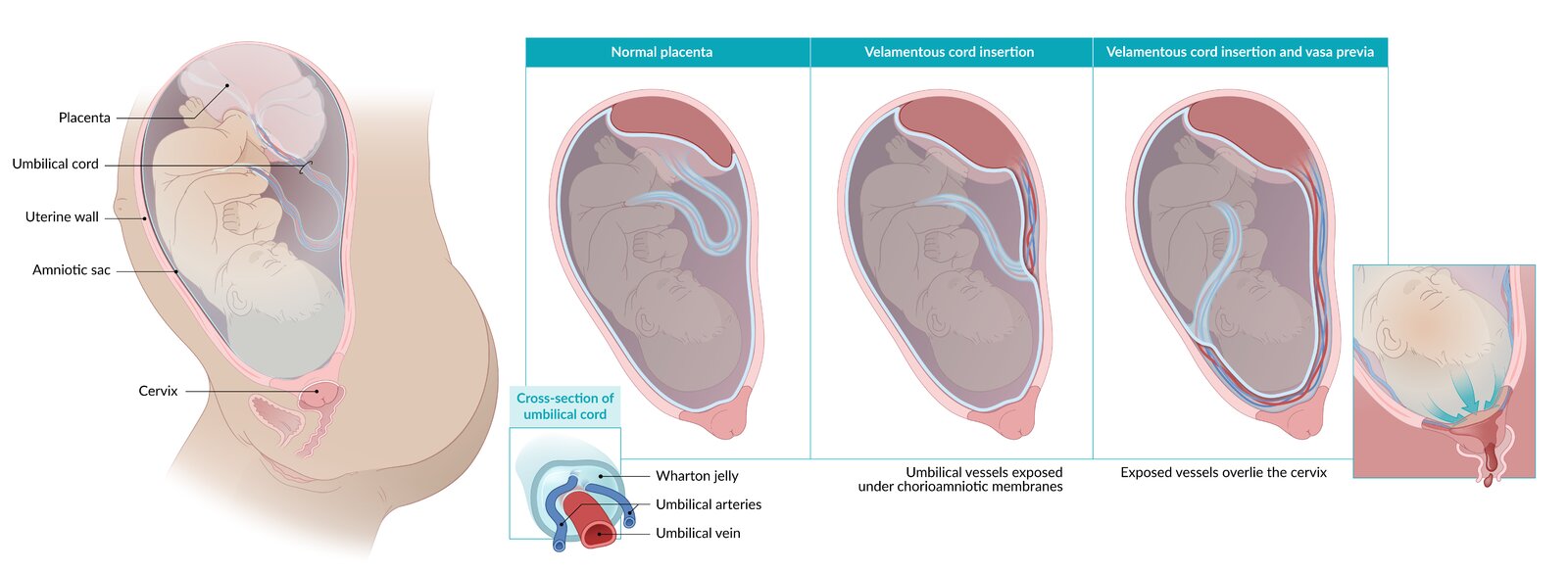
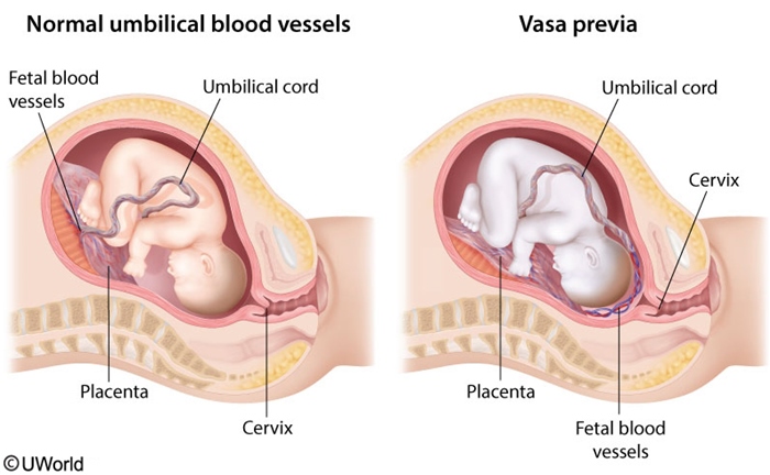
Vasa previa is a rare but life-threatening obstetric condition in which unprotected fetal vessels traverse the fetal membranes across or near the internal cervical os, placing them at risk of rupture during labor or membrane rupture. These exposed vessels are covered only by the thin chorionic and amniotic membranes, lacking the protective Wharton jelly that normally surrounds the umbilical cord.
It is the only cause of antepartum hemorrhage of purely fetal origin. The estimated incidence is approximately 1 in 2,500 births, though the true prevalence may be higher in pregnancies with certain risk factors (e.g., in vitro fertilization, where incidence approaches 1 in 25–75).
Etiology and Pathophysiology
Vasa previa arises from abnormal placental or umbilical cord insertion. The two most common anatomic defects are:
- Velamentous insertion of the umbilical cord — fetal vessels insert directly into the fetal membranes instead of the placental disk, leaving them exposed to the intrauterine environment rather than protected within the umbilical cord.
- Succenturiate or bilobate placenta — a separated accessory placental lobe connected to the main placenta by fetal vessels that traverse the membranes between the lobes, crossing the internal cervical os.
Without Wharton jelly protection, these membraneous vessels are highly vulnerable to rupture when membranes rupture or during cervical dilation in labor. Hemorrhage originates from the fetoplacental circulation; because fetal blood volume is only ~250 mL at term, even modest blood loss (20–50 mL) rapidly leads to fetal anemia, cardiovascular collapse, and exsanguination. The fetal heart rate response to acute anemia produces the characteristic sinusoidal pattern (slow oscillations of 2–3 Hz baseline with absent variability) on the cardiotocogram.
Risk Factors
Three primary risk factors account for the vast majority of vasa previa cases and should be prioritized in antenatal screening:
| Risk Factor | Pathophysiologic Basis | |
| Placental anomalies | Velamentous insertion or succenturiate/bilobate placenta | Vessels insert directly into fetal membranes instead of placental disk, leaving them unprotected by Wharton jelly |
| Placenta previa or low-lying placenta | Abnormally low placental position with vessels crossing internal os | May coexist with velamentous insertion; increased risk in multiple gestations and IVF |
| In vitro fertilization | Assisted reproductive technology | 2–3× increased incidence; exact mechanism unclear but may relate to abnormal trophoblastic invasion and placental development |
Note — Although multiparity and advanced maternal age are listed in some sources as minor risk factors, they are much lower-yield than the three above and should not guide screening strategy.
Clinical Features
The classic presentation of vasa previa is a triad of clinical signs, almost always triggered by rupture of membranes:
| Clinical Feature | Description | |
| Painless vaginal bleeding | Bright-red fetal blood loss (not maternal) | Often occurs immediately after spontaneous or iatrogenic rupture of membranes; caused by tearing of exposed fetal vessels. In ~25% of cases, bleeding may occur without ROM due to vessel compression or spontaneous microrupture. |
| Acute fetal distress | Bradycardia, variable decelerations, sinusoidal FHT | Sinusoidal pattern (2–3 Hz baseline oscillations with absent beat-to-beat variability) is pathognomonic for fetal anemia from acute hemorrhage. This is NOT the same as sinusoidal-like patterns from other causes (e.g., methadone, fetal hydrops). |
| Rapid progression to fetal compromise | Fetal shock, bradycardia, loss of cardiac output | Deterioration occurs within minutes to a few hours if left untreated. Because fetal blood volume is only ~250 mL, loss of just 50–100 mL (~20–40%) causes profound anemia and cardiovascular collapse. |
| Vasa Previa Presentation: BRD | |
|
B = Bleeding (painless, fetal origin) Memory aid: ROM triggers BRD in vasa previa—the only fetal hemorrhage emergency. |
جملة تذكرية |
| Important – فخ امتحاني | |
|
The bleeding in vasa previa is strictly fetal blood, not maternal blood. This causes rapid fetal bradycardia and a sinusoidal pattern on the CTG (from acute anemia). In contrast, placenta previa presents with maternal vaginal bleeding, and the fetus is typically stable initially (normal fetal heart rate). |
Diagnosis
Antenatal diagnosis is crucial and markedly improves perinatal outcomes by allowing planned, elective cesarean delivery before labor or ROM occurs. Postpartum diagnosis—made by examining the placenta and membranes for vessels crossing the cervix—is too late to prevent fetal death.
Ultrasound with color Doppler (transvaginal or transabdominal) is the primary diagnostic tool. Findings include:
- Fetal blood vessels coursing directly across the internal cervical os (seen best in the sagittal plane on transvaginal ultrasound)
- Color flow in these vessels (red or blue) indicating blood flow direction (usually from umbilical arteries or a vein connecting lobes of the placenta)
- Absence of Wharton jelly surrounding the crossing vessels (they are covered only by thin chorionic and amniotic membrane)
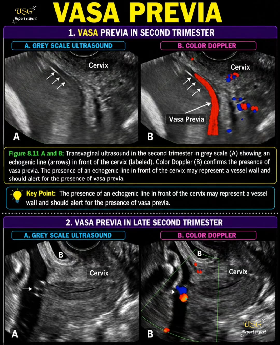
Screening indications: All pregnancies should be screened for vasa previa at the 18–24 week anatomy scan, with particular vigilance in pregnancies with known risk factors (IVF conception, placenta previa, velamentous insertion detected on imaging, multiple gestation).
| Important – ملاحظة سريرية | |
|
Always look for keywords in the ultrasound report: "velamentous insertion," "succenturiate lobe," "bilobate placenta," or "vessels crossing the internal os." If you see these descriptors in a patient presenting with painless bleeding after ROM and fetal distress, vasa previa is confirmed. Do not miss these in the imaging report—they are the diagnostic clues that allow life-saving antenatal intervention. |
Management
Management is stratified by whether vasa previa is diagnosed before or during labor:
| Management Strategy by Clinical Scenario | |||
|---|---|---|---|
| Scenario | Diagnosis Timing | Intervention | Timing |
| Emergency cesarean delivery | Diagnosed in active labor or after ROM with fetal distress | Immediate emergency cesarean delivery (category 1 urgency); do not delay for any reason | Within minutes of diagnosis; fetal survival depends on this |
| Planned cesarean delivery | Diagnosed antenatally on screening ultrasound | Hospitalization for close fetal monitoring; planned cesarean delivery before onset of labor or spontaneous ROM | At 34–37 weeks (balancing prematurity risk against hemorrhage risk); delivery must be elective, not induced in labor |
Detailed management approach:
- Antenatal diagnosis: Once vasa previa is confirmed on ultrasound, admit the patient for hospital admission (or at least daily monitoring) at ~32–34 weeks. Some centers recommend inpatient admission even earlier (e.g., 30 weeks) depending on local resources.
- Fetal monitoring: Twice-daily cardiotocography (NST) to detect early fetal distress; educate patient to report any vaginal bleeding or symptoms of ROM immediately.
- Planned delivery: Perform elective cesarean delivery at 34–37 weeks (exact timing depends on lung maturity risk vs. hemorrhage risk; most centers deliver at 34–36 weeks). Do not attempt vaginal delivery.
- Peripartum care: Have neonatal resuscitation and transfusion capability available at delivery; prepare for possible neonatal anemia if fetal bleeding occurs during surgery.
| Important – ملاحظة سريرية | |
|
If the patient presents with ruptured membranes and fetal distress (tachycardia/sinusoidal pattern + painless bleeding), the only correct answer is immediate emergency cesarean section. |
Key Points for Exams – نقاط مهمة للامتحانات
- Vasa previa is the only cause of purely FETAL antepartum hemorrhage. Distinguish it from placenta previa (maternal bleeding) by the presence of fetal distress and sinusoidal FHT pattern immediately after ROM.
- Classic triad (BRD): Painless vaginal bleeding triggered by ROM, acute fetal distress (bradycardia + sinusoidal pattern), rapid progression to fetal compromise if untreated.
- Diagnosis is ultrasound-based: Transvaginal color Doppler showing fetal vessels crossing the internal cervical os WITHOUT surrounding Wharton jelly. Antenatal diagnosis is life-saving.
- Two anatomic defects account for 90% of cases: Velamentous insertion of the umbilical cord and succenturiate/bilobate placenta. Both leave fetal vessels exposed to the vaginal environment.
- High-risk groups requiring screening: IVF conception (~3% incidence), placenta previa or low-lying placenta, multiple gestation, dichorionic diamniotic twins.
- Management is stratified: Antenatal diagnosis → elective cesarean at 34–37 weeks (prevents hemorrhage entirely); diagnosis in labor or after ROM → emergency cesarean delivery within minutes (survival depends on this).
- Exam pearl: If a CTG shows a sinusoidal pattern in a patient with painless vaginal bleeding after ROM, suspect fetal hemorrhage first (vasa previa) before other causes of sinusoid-like patterns.
- Never attempt vaginal delivery in a pregnancy diagnosed with vasa previa. The risk of vessel rupture during labor and cervical descent is unacceptable.
احصل على التجربة الكاملة
اشترك للوصول لفيديوهات الشرح التفصيلي والبطاقات التعليمية التفاعلية وأسئلة الممارسة مع تتبع التقدم.