Summary
Placenta accreta spectrum is abnormal placental invasion into the uterus, most often linked to prior cesarean section or placenta previa. It may cause failure of placental separation and severe hemorrhage, with management usually by planned cesarean hysterectomy.
Early antenatal diagnosis and delivery at a tertiary center are critical for reducing maternal morbidity and mortality.
Definition & Classification
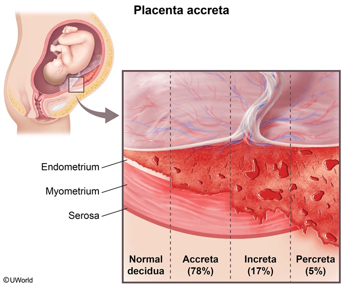
Placenta accreta spectrum (PAS) refers to abnormal placental adherence and invasion into the uterine wall, representing a spectrum of disorders classified according to the depth of trophoblastic invasion. Normally, placental villi penetrate only the superficial decidua basalis. When this boundary is breached, varying degrees of abnormal invasion occur, ranging from simple attachment to complete penetration.
| AIP Mnemonic – جملة تذكرية | |
|
Attaches (Accreta) | Invades (Increta) | Penetrates (Percreta) Remember: each letter describes the progressive depth of abnormal trophoblastic invasion. |
جملة تذكرية |
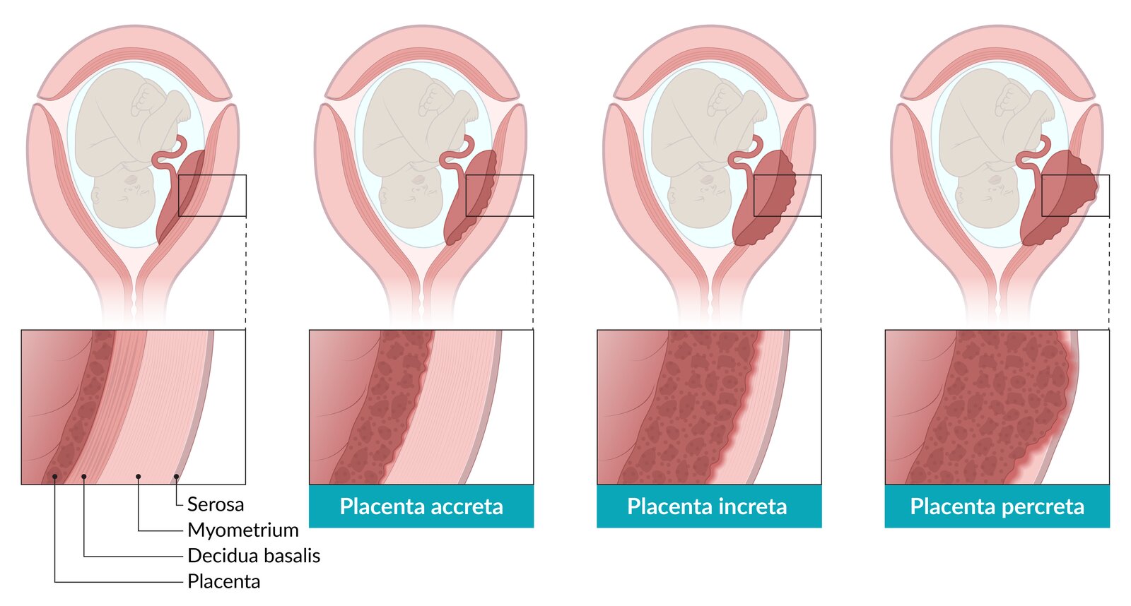
The three types are distinguished by the depth of invasion into the uterine wall:
| Classification of Placenta Accreta Spectrum: By Depth of Trophoblastic Invasion | |
| Placenta Accreta | Attachment to Myometrium |
| Pathology | Abnormal villous attachment directly to myometrium without penetration; decidua basalis is absent or defective |
| Frequency | ~63% of all PAS cases |
| Clinical Impact | May still cause severe PPH due to failure of normal separation; hysterectomy usually required |
| Placenta Increta | Invasion into Myometrium |
| Pathology | Villous invasion extends into (but not through) the myometrium |
| Frequency | ~15% of all PAS cases |
| Clinical Impact | Significant hemorrhage risk; hysterectomy often necessary |
| Placenta Percreta | Penetration Through Myometrium |
| Pathology | Villi penetrate entire myometrium and visceral peritoneum; may invade bladder, bowel, or other adjacent organs |
| Frequency | ~22% of all PAS cases |
| Clinical Impact | Highest morbidity/mortality; risk of bladder hematuria, ureteral injury, and life-threatening hemorrhage |
| Exam Pearl – فخ امتحاني | |
|
Don't confuse the types. Accreta just attaches to the myometrium, Increta goes into it, and Percreta goes through it—sometimes hitting the bladder. If you see hematuria in the third trimester with a prior cesarean, think Percreta (bladder invasion). |
تذكر |
The anatomical illustrations below demonstrates the spectrum of abnormal placental attachment:
Risk Factors
Placenta accreta spectrum is strongly associated with uterine scarring and abnormal placental implantation. The risk is proportional to the degree and extent of disruption to the normal decidualization process. Three risk factors account for the vast majority of cases and are heavily tested on board exams:
| Major Risk Factors for Placenta Accreta Spectrum | |
| 1. Previous Cesarean Section | Most Critical Risk |
| Mechanism | Uterine scarring creates defective decidualization; risk increases with each additional cesarean delivery |
| Exam Relevance | Present in >90% of PAS cases; always screen for prior CS history in third-trimester complications |
| 2. Placenta Previa (Especially Over a Scar) | High-Yield Board Combo |
| Synergistic Effect | Placenta implanting over a previous uterine incision dramatically increases abnormal invasion risk |
| Exam Relevance | This combination (prior CS + placenta previa) is the classic board presentation |
| 3. Prior Uterine Surgery or Curettage | Additional Scar |
| Includes | Dilation and curettage (D&C), myomectomy, metroplasty, or hysteroscopic procedures |
| Exam Relevance | Any disruption of the normal endometrial-myometrial interface increases PAS risk |
| Clinical Pearl – ملاحظة سريرية | |
|
The most high-yield risk factor combination for the board exam is a patient with placenta previa who has a history of multiple previous cesarean sections. Always screen for this in your third-trimester differential. |
ملاحظة |
Clinical Features
Placenta accreta spectrum is often asymptomatic during pregnancy and may be detected incidentally on routine antenatal ultrasound. However, when symptoms do occur, they are often ominous. Key clinical features include:
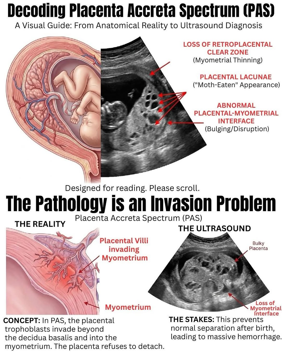
- Ultrasound findings (antenatal): Loss of the normal hypoechoic retroplacental zone, irregular placental lacunae ("Swiss cheese" appearance), increased vascularity on color Doppler, and abnormal intraplacental vascularity.
- Third-trimester vaginal bleeding: Occurs occasionally and may be the first clue to placental abnormality, especially when associated with prior cesarean.
- Failure of placental separation at delivery: The placenta remains firmly adherent to the uterine wall after delivery of the infant. Normal mechanisms of placental detachment (uterine contraction, traction on the umbilical cord) fail. This is followed by massive postpartum hemorrhage, with average blood loss of 3,000–5,000 mL or higher. Many patients require transfusion of multiple units of packed red blood cells, fresh frozen plasma, and platelets. Without prompt hysterectomy, exsanguination and maternal death may occur.
Diagnosis
Diagnosis of placenta accreta spectrum relies primarily on imaging, though many cases remain undiagnosed until labor or delivery.
Ultrasound is the first-line imaging modality and should be performed in any pregnant patient with risk factors (prior cesarean + third-trimester bleeding, or placenta previa). Key diagnostic features include:
- Loss of hypoechoic decidual layer
- Irregular placental lacunae
- Abnormal vascularity crossing placental-myometrial interface
- Placental bulging into bladder or adjacent organs
MRI may be helpful in select cases where ultrasound is inconclusive, particularly in patients with posterior placentation or maternal obesity, or when percreta with bladder invasion is suspected.
Important limitation: Many cases of PAS remain completely undiagnosed until labor or delivery, when the placenta fails to separate despite maternal uterine contractions and traction on the umbilical cord. This intrapartum discovery often coincides with massive hemorrhage and necessitates emergency hysterectomy.
Management
| Management of Placenta Accreta Spectrum | ||
|---|---|---|
| Antenatally Diagnosed | ||
| Timing | Setting | Key Actions |
| Planned cesarean delivery (typically 36–37 weeks, after fetal lung maturity assessment) | Tertiary care center with multidisciplinary team (OB, anesthesia, ICU, blood bank, interventional radiology) | Scheduled cesarean hysterectomy with placenta left in situ; prepare for massive transfusion protocol; consider prophylactic balloon catheters or interventional radiology backup |
| Undiagnosed Until Delivery | ||
| Scenario | Immediate Response | Surgical Decision |
| Placenta does not separate after vaginal delivery or low-transverse cesarean | Do NOT forcefully pull on the cord. Activate massive transfusion protocol; call for emergent surgical backup and anesthesia | Prepare for urgent cesarean hysterectomy with a goal to minimize blood loss and prevent maternal exsanguination |
| Conservative Management (Select Cases) | ||
| Indication | Approach | Risks |
| Only considered in hemodynamically stable patients with partial placental retention and no ongoing hemorrhage | Leave placenta in situ; close uterus; close skin; plan for close outpatient follow-up or readmission for imaging/ultrasound | High risk of infection (chorioamnionitis, endomyometritis), delayed hemorrhage (days to weeks later), need for emergency hysterectomy, and potential loss of fertility |
| Clinical Note – ملاحظة سريرية | |
|
In the operating room: If the placenta doesn't separate after delivery and you see massive hemorrhage, do NOT keep pulling on the cord. You need to prepare for a rapid cesarean hysterectomy and activate your massive transfusion protocol immediately. Time is critical. |
ملاحظة |
Key Points for Exams – نقاط مهمة للامتحانات
- Classic presentation: Placenta previa + history of one or more prior cesarean sections = high suspicion for PAS. Always ask about prior uterine surgery in the third-trimester bleeding differential.
- Risk increases with each cesarean: A patient with three prior CS carries significantly higher risk than a patient with one prior CS.
- Antenatal diagnosis is crucial: Planned delivery at 36–37 weeks at a tertiary center with cesarean hysterectomy reduces maternal mortality compared to intrapartum discovery.
- The three types (AIP mnemonic): Accreta attaches to myometrium (~63%); Increta invades into it (~15%); Percreta penetrates through it (~22%). Each carries risk of severe PPH.
- Percreta red flag: If a patient in the third trimester with prior CS develops hematuria, suspect bladder invasion (percreta). This requires preoperative imaging and surgical planning to avoid ureteral injury.
- Ultrasound findings: Loss of retroplacental lucency, irregular lacunae (Swiss cheese appearance), and increased vascularity on Doppler are key diagnostic features. MRI is used in select cases.
- Massive transfusion protocol: Patients with PAS require pre-delivery preparation for >5 units of pRBC, FFP, and platelets. Deliver at a tertiary center with ICU support.
- Intrapartum red flag: Placenta that does NOT separate after delivery despite strong contractions + traction on cord = prepare for emergency hysterectomy. Do not force placental separation; activate massive transfusion protocol and call for surgical backup immediately.
- Conservative management is rare: Leaving the placenta in situ carries high risk of infection, delayed hemorrhage, and loss of fertility. Reserved only for stable, selected cases.
- Maternal morbidity and mortality: Even with planned delivery, morbidity includes massive transfusion (up to 40% of cases), ICU admission, and hysterectomy (with loss of future pregnancy capacity). Fetal and maternal mortality are reduced significantly with antenatal diagnosis.
احصل على التجربة الكاملة
اشترك للوصول لفيديوهات الشرح التفصيلي والبطاقات التعليمية التفاعلية وأسئلة الممارسة مع تتبع التقدم.