SUMMARY
Secondary postpartum hemorrhage (PPH) is abnormal or excessive vaginal bleeding occurring 24 hours to 12 weeks after delivery, most often in the second postpartum week. Its four major causes are remembered as the 4 Ts: Tissue (retained products of conception), Tone (subinvolution of the placental site), Temperature (postpartum endometritis), and Thrombin (coagulation disorders).
Diagnosis combines clinical assessment, pelvic ultrasound with color Doppler, and laboratory studies (CBC, coagulation profile, cultures). After stabilization, management is cause-specific — uterotonics, uterine evacuation, IV antibiotics, or blood/factor replacement — with embolization, artery ligation, or hysterectomy reserved for refractory, severe bleeding.
Overview
Definition
Secondary PPH is abnormal or excessive vaginal bleeding occurring between 24 hours and 12 weeks after delivery. It is less common than primary PPH (<24 hours) but can be life-threatening if it is not recognized and treated quickly.
Epidemiology
- Peak timing: second week postpartum.
- Most common causes: endometritis and retained products of conception (RPOC).
- Subinvolution of the placental site: ~13% of cases.
Etiology – The 4 Ts of Secondary PPH
Secondary PPH has four main causes, remembered as the 4 Ts. Three are uterine (Tissue, Tone, Temperature). They overlap clinically and are told apart mainly by risk factors and ultrasound/Doppler pattern. The fourth (Thrombin) is systemic. Each cause is covered in full in the sections that follow.
| Mnemonic – جملة تذكرية: The 4 Ts of Secondary PPH | |
جملة تذكرية: بقايا – ارتخاء – حرارة – تجلّط |
جملة تذكرية |
Rare causes:
- Disseminated intravascular coagulation (DIC): an acquired coagulopathy, covered under Thrombin.
- Subacute or chronic uterine inversion (presenting >24 hours after delivery).
See the uterine inversion pathophysiology, presentation, and management summary for its causes and corrective steps.
Tissue & Tone – RPOC and Subinvolution of the Placental Site
1. Retained Products of Conception (RPOC): "Tissue"
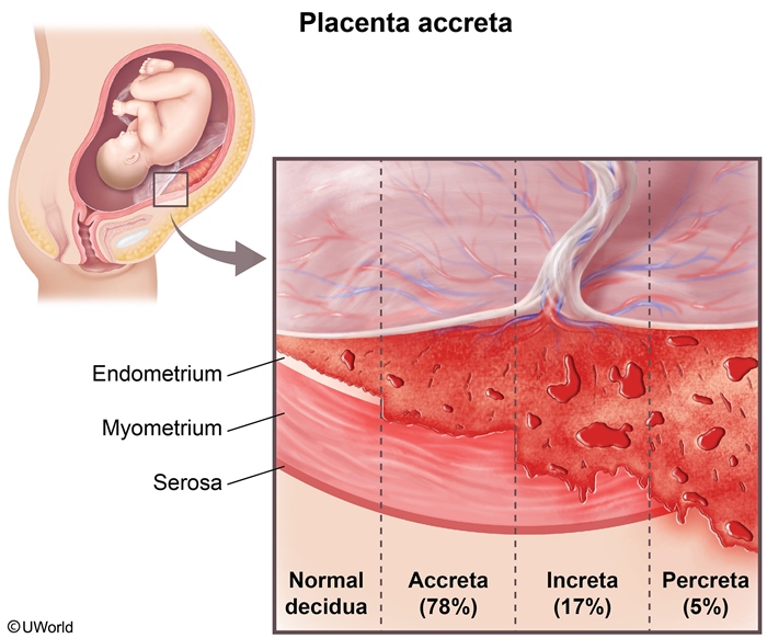
Risk factors: placenta accreta spectrum, retained placenta, preterm birth, multiple gestation, and assisted (operative) vaginal delivery.
In placenta accreta spectrum, the placenta invades abnormally into the myometrium. Placental tissue therefore cannot separate completely:
Clinical features: abnormal uterine bleeding, lower abdominal pain, ± fever, amenorrhea. On examination the uterus is enlarged.
| Important – فكرة سؤال | |
|
Intraamniotic infection predisposes to RPOC even when it was adequately treated — intrauterine inflammation makes the placental membranes adhere to the uterine wall. Unlike a retained placenta (immediate PPH with a boggy uterus), RPOC causes late PPH with a boggy OR firm uterus. A firm uterus does not exclude RPOC — the pelvic ultrasound decides. |
تذكر |
Imaging: pelvic ultrasound with color Doppler (first-line). Look for a thickened echogenic endometrial complex or a focal intrauterine mass. Vascularity within the endometrial material on color Doppler is the key finding.
Management:
- Step 1 – Medical: misoprostol (PGE1 analog) 600–1,000 mcg PO, SL, or PR as a single dose, for a stable patient with modest bleeding.
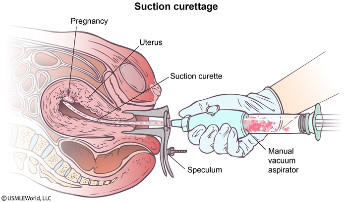
- Step 2 – Surgical: dilation and curettage (suction curettage) or hysteroscopic removal, if bleeding is heavy or tissue persists after medical therapy. Avoid overly aggressive curettage of the postpartum uterus, which risks Asherman syndrome.
2. Subinvolution of the Placental Site: "Tone"
Risk factors: multiparity, cesarean delivery, uterine atony, endometritis, coagulopathy, and RPOC.
Clinical features: heavy bleeding, classically in the second week postpartum, ± fever, abdominal pain, and signs of hypovolemia.
Imaging: ultrasound shows hypoechoic, tortuous myometrial vessels. Doppler shows an increased peak systolic velocity.
Histology (confirmatory / gold standard, rarely required): dilated myometrial arteries with thickened walls and thrombosis.
| ملاحظة – Note | |
|
Exam pitfall: To differentiate RPOC from subinvolution on color Doppler, always identify where vascularity is located. If vascularity is within the endometrial material, RPOC is certain; conversely, if you identify hypoechoic tortuous vessels within the myometrium with elevated peak systolic velocity, subinvolution is the diagnosis. |
ملاحظة |
Management:
- Step 1 – Uterotonic: oxytocin 10–40 units in 500–1,000 mL crystalloid as a continuous IV infusion.
- Step 2 – Evacuation: D&C or vacuum removal, if bleeding continues despite uterotonics.
- Step 3 – Severe or refractory bleeding: uterine artery embolization (fertility-sparing) or hysterectomy.
Refer to the uterotonics comparison for the mechanisms of oxytocin and misoprostol.
Temperature – Postpartum Endometritis
3. Temperature – Postpartum Endometritis
Risk factors: cesarean delivery (strongest), prolonged rupture of membranes, prolonged labor, multiple cervical examinations, meconium-stained amniotic fluid, retained tissue, and low socioeconomic status. The risk factors for postpartum endometritis table adds intraamniotic infection, GBS colonization, bacterial vaginosis, and manual placental removal.
Clinical features: fever (typically >24 hours postpartum), chills, malaise, lower abdominal pain, uterine (fundal) tenderness, and foul-smelling lochia. Bleeding may be increased.
Diagnosis: mainly clinical, supported by cultures.
Serum: blood cultures, CBC.
Urine: urine culture.
Vaginal discharge: culture. The infection is typically polymicrobial.
Imaging: ultrasound is not needed to make the diagnosis. Use it only to detect coexisting RPOC.
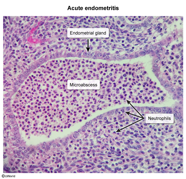
On histology, acute endometritis shows neutrophils infiltrating the endometrial glands and stroma:
Management:
- Step 1 – IV antibiotics: clindamycin 900 mg IV every 8 hours + gentamicin 5 mg/kg IV every 24 hours (or 1.5 mg/kg every 8 hours). Continue until the patient has been afebrile and asymptomatic for 24–48 hours.
- Step 2 – Uterine evacuation: if RPOC is present.
- Step 3 – Hysterectomy: for refractory, life-threatening infection.
| ملاحظة – Note | |
|
Clinical note: Once the patient becomes afebrile on IV clindamycin + gentamicin, there is no need to continue oral antibiotics. If fever persists despite antibiotics, perform an ultrasound and search for RPOC because it requires evacuation. |
ملاحظة |
The postpartum endometritis risk factors, clinical features, etiology, and treatment table summarizes the full picture on one page.
Thrombin – Coagulation Disorders and DIC
4. Thrombin – Coagulation Disorders and DIC
Causes:
- Acquired: HELLP syndrome, sepsis, intrauterine fetal demise, and disseminated intravascular coagulation (DIC). DIC is typically triggered by placental abruption, preeclampsia, amniotic fluid embolism, or prolonged fetal demise.
- Inherited: von Willebrand disease, hemophilia, and other factor deficiencies (e.g., factor XIII).
Refer to the HELLP syndrome clinical features and laboratory findings, the abruptio placentae overview, the amniotic fluid embolism summary, and the intrauterine fetal demise evaluation and management for the details of each trigger.
Clinical clues:
Mucocutaneous bleeding, GI bleeding, a history of menorrhagia, and oozing from wounds or IV sites. Bleeding continues even though the uterus is firm and contracted.
| Important – فكرة سؤال | |
|
von Willebrand disease classically presents as secondary (delayed) PPH. Estrogen raises vWF during pregnancy and masks the defect; levels fall after delivery → bleeding days to weeks later. Clue: small, firm, nontender uterus with a normal ultrasound (no mass, no increased vascularity) yet persistent bleeding. Labs: normal platelets and PT, aPTT normal or prolonged, prolonged bleeding time. Treat acute bleeding with desmopressin. |
تذكر |
Diagnosis:
Serum (first-line screen): CBC (platelets), PT, aPTT, fibrinogen, D-dimer.
Serum (suspected inherited disorder): vWF antigen and vWF activity ± factor VIII level.
Typical DIC pattern:
- Platelets: ↓
- PT / aPTT: prolonged
- Fibrinogen: ↓
- D-dimer: ↑
The laboratory characteristics of coagulopathies table compares the platelet, PT, PTT, and fibrinogen patterns of vWD and DIC. See the laboratory characteristics of disseminated intravascular coagulation (DIC) for its triggers, mechanism, and full laboratory profile, including schistocytes.
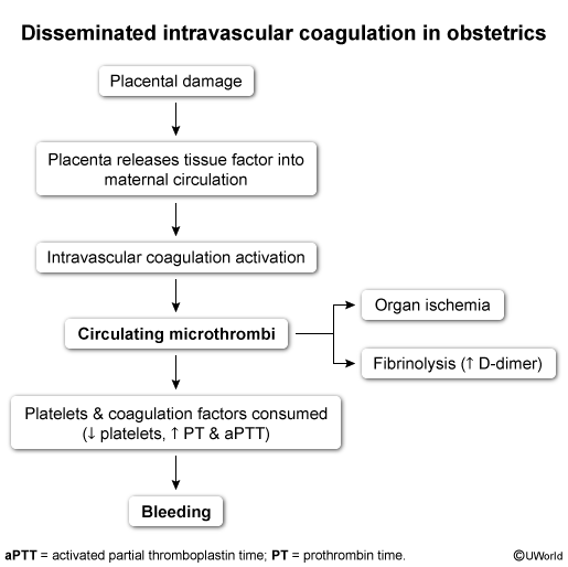
In obstetric DIC, placental damage releases tissue factor. This drives consumptive coagulopathy and paradoxical bleeding:
Management:
- Blood products: FFP 10–15 mL/kg IV. Transfuse platelets for thrombocytopenia with active bleeding.
- Factor replacement: specific concentrates (e.g., vWF-containing concentrate, factor VIII/IX, factor XIII).
- Desmopressin: 0.3 mcg/kg IV over 15–30 minutes. It releases endothelial vWF (type 1 vWD, mild hemophilia A).
- Antifibrinolytic (tranexamic acid): 1 g IV over 10 minutes. Repeat 1 g if bleeding continues after 30 minutes.
- DIC: ICU care, removal of the inciting cause, and selective blood product replacement.
See the effects of desmopressin (DDAVP) therapy for how it raises vWF and factor VIII levels.
Clinical Features, Diagnosis & Management Principles
Clinical Features
- Abnormal, often heavy or recurrent vaginal bleeding after the first 24 hours postpartum.
- May be associated with fever, lower abdominal pain, uterine tenderness, and signs of anemia or hypovolemia.
Use these red flags to separate it from physiologic lochia:
- Clots: passage of large clots
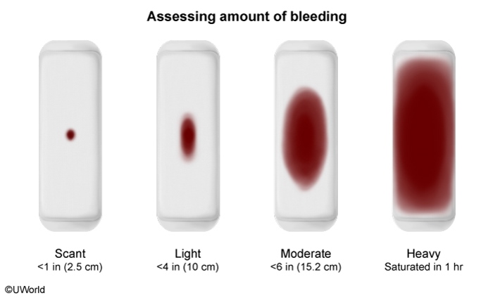
- Pad count: saturation of ≥1 pad/hour for ≥2 consecutive hours
- Symptoms: symptomatic anemia (dizziness, chest pain) or hemodynamic instability
Pad saturation gives a bedside estimate of bleeding volume:
See the normal postpartum lochia types and duration for the expected rubra → serosa → alba timeline that serves as the baseline.
| Important – فكرة سؤال | |
Normal lochia can transiently increase at 7–14 days postpartum when the eschar over the placental site sloughs (heavier bleeding lasting <2 hours). Small clots, stable vitals, a firm nontender uterus, and an appropriate hemoglobin = observation and reassurance — not D&C or antibiotics. |
تذكر |
Diagnosis
Clinical evaluation: bleeding severity (pad count, clots, vital signs) and systemic symptoms (fever, pain, uterine tenderness).
Serum: CBC and coagulation profile (PT, aPTT, fibrinogen) in every patient. Add blood cultures when infection is suspected.
Imaging: pelvic ultrasound ± color Doppler (first-line) for RPOC or subinvolution.
Histopathology: rarely required, but confirmatory for subinvolution.
See the differential diagnosis of postpartum hemorrhage for the examination findings that help localize the cause at the bedside.
Management Principles
- Step 1 – Stabilization: resuscitate with IV fluids, and give blood products if there is hemodynamic compromise or significant anemia.
- Step 2 – Identify the "T": CBC, coagulation profile, cultures if febrile, and pelvic ultrasound ± Doppler.
- Step 3 – Targeted treatment (full regimens are in each cause's section):
- Endometritis: IV antibiotics (clindamycin + gentamicin) ± uterine evacuation.
- RPOC: uterotonic drugs (misoprostol), D&C, or hysteroscopic removal.
- Subinvolution: uterotonics (oxytocin), surgical evacuation, embolization, or hysterectomy in severe cases.
- Coagulopathy: factor replacement, desmopressin, and transfusion support.
- Rare causes (DIC, inversion): specific corrective measures with ICU support. For DIC, remove the trigger and replace blood products selectively. For inversion, reposition the uterus.
- Step 4 – Refractory or unexplained bleeding (no correctable cause found): uterine artery embolization, uterine or internal iliac artery ligation, and hysterectomy as the last resort.
Severe hemorrhage with hypotension can infarct the pituitary. Refer to the Sheehan syndrome table for the resulting hormone deficiencies (failure of lactation, amenorrhea, secondary adrenal insufficiency).
Key Points for Exams – نقاط مهمة للامتحانات
- Definition: secondary PPH is bleeding from 24 hours to 12 weeks postpartum, peaking in week 2.
- Most common causes: endometritis and RPOC. Subinvolution accounts for ~13%.
- 4 Ts: Tissue (RPOC), Tone (subinvolution), Temperature (endometritis), Thrombin (coagulopathy). Rare causes: DIC and subacute/chronic uterine inversion.
- Red flags vs normal lochia: large clots, ≥1 pad/hour for ≥2 consecutive hours, symptomatic anemia. A brief increase in bleeding at 7–14 days (eschar sloughing) is physiologic.
- Doppler: vascularity within the endometrial material → RPOC. Hypoechoic tortuous myometrial vessels with ↑ peak systolic velocity → subinvolution.
- RPOC: risk factors include placenta accreta spectrum, retained placenta, and prior intraamniotic infection. The uterus may be boggy or firm. Treat with misoprostol (PGE1 analog) → D&C or hysteroscopic removal. Aggressive curettage risks Asherman syndrome.
- Subinvolution: confirmed by histology (dilated, thick-walled, thrombosed myometrial arteries), though this is rarely needed. Treat with IV oxytocin → D&C → embolization or hysterectomy.
- Endometritis: fever + fundal tenderness + foul-smelling lochia. Cesarean delivery is the strongest risk factor. Treat with IV clindamycin + gentamicin. If fever persists despite antibiotics, look for RPOC and evacuate.
- Coagulopathy clue: small, firm uterus + normal ultrasound + persistent bleeding → think von Willebrand disease. Order vWF antigen/activity and treat with desmopressin.
- DIC: triggered by abruption, preeclampsia, amniotic fluid embolism, or prolonged fetal demise. Labs show ↓ platelets, ↓ fibrinogen, prolonged PT/aPTT, ↑ D-dimer. Management is ICU care, removing the trigger, and selective blood product replacement.
- Approach: stabilize → CBC, coagulation profile, ± cultures → pelvic ultrasound ± Doppler → treat the identified "T."
- Refractory bleeding: uterine artery embolization or uterine/internal iliac artery ligation, with hysterectomy as the last resort.
- Long-term sequela: severe hemorrhage with hypotension → Sheehan syndrome (failure of lactation, amenorrhea).
احصل على التجربة الكاملة
اشترك للوصول لفيديوهات الشرح التفصيلي والبطاقات التعليمية التفاعلية وأسئلة الممارسة مع تتبع التقدم.