شرح المدرسين

SUMMARY
Rh isoimmunisation is a preventable cause of severe fetal and neonatal morbidity. Advances in universal maternal screening, routine anti-D prophylaxis, and modern fetal monitoring (MCA Doppler) have dramatically reduced its incidence and improved outcomes. While other causes of HDFN (ABO, Kell) remain clinically relevant, strict adherence to anti-D immunoglobulin protocols remains the most effective intervention in safeguarding against this condition.
DEFINITION & PATHOPHYSIOLOGY
Rh isoimmunisation (erythroblastosis fetalis) refers to maternal alloimmunisation against the fetal Rhesus D (RhD) antigen, causing haemolytic disease of the fetus and newborn (HDFN). Severity ranges from mild neonatal jaundice to severe fetal anaemia, hydrops fetalis, and intrauterine death.
Pathophysiology
Isoimmunisation begins when fetal RBCs enter the maternal circulation during sensitising events:
- Delivery, miscarriage, abortion
- Antepartum haemorrhage or trauma
- Invasive obstetric procedures (amniocentesis, chorionic villus sampling)
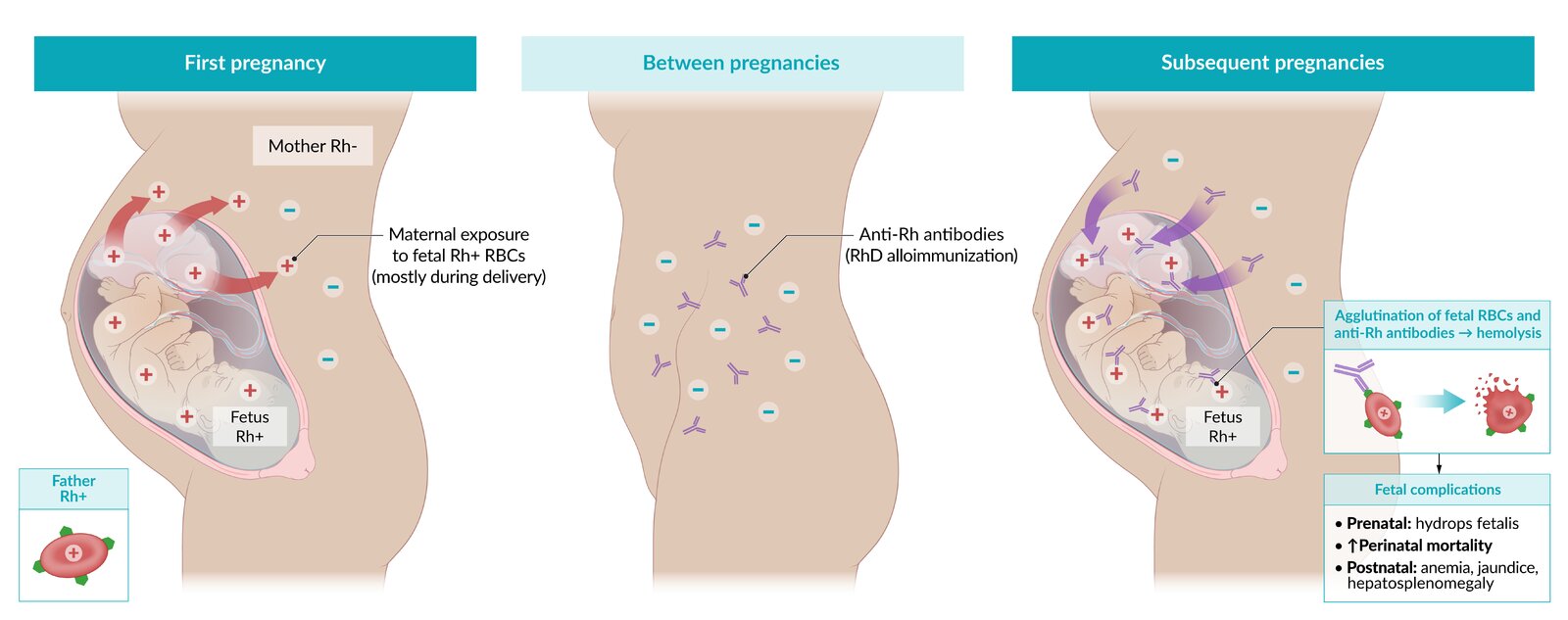
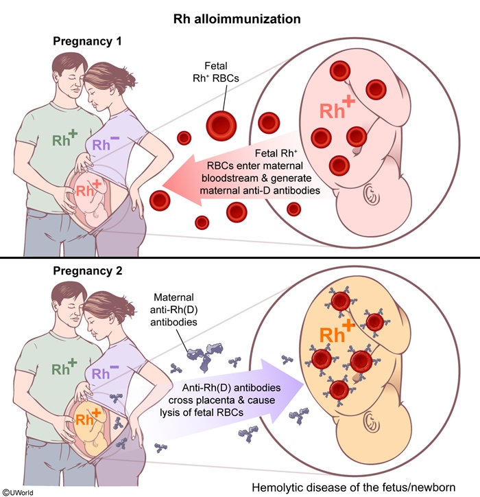
In an unsensitised RhD-negative woman carrying an RhD-positive fetus, maternal exposure triggers two immune phases:
- Primary response (first pregnancy): IgM anti-D antibodies are produced. IgM does NOT cross the placenta → the first fetus is typically spared.
- Secondary response (subsequent pregnancies): Memory B-cells rapidly produce IgG anti-D, which does cross the placenta, bind to fetal erythrocytes, and cause haemolysis.
| فخ امتحاني – Clinical Pearl | |
|
The first RhD-incompatible pregnancy is usually unaffected because the primary response generates IgM, which remains in the maternal circulation and does not cross the placental barrier. Only in second and subsequent pregnancies does the rapid secondary response of IgG cause fetal haemolysis. This is a classic exam distinction and differentiates Rh incompatibility from ABO incompatibility (which can affect the first pregnancy). |
تذكر |
Haemolysis leads to fetal anaemia, extramedullary haematopoiesis, hepatosplenomegaly, hydrops fetalis, hyperbilirubinaemia, and in severe cases kernicterus or intrauterine death.
Other blood group incompatibilities: While RhD is most severe, Kell alloimmunisation is the second-leading cause of severe HDFN (causes both haemolysis and erythropoiesis suppression). ABO incompatibility is far more common (~20% of pregnancies) but usually mild.
CLINICAL FEATURES
Prenatal Manifestations
Hydrops fetalis (in severe cases):
- Ascites, pleural/pericardial effusion, skin oedema, placental thickening
- Cardiomegaly, increased cardiothoracic ratio
- Polyhydramnios (from fetal heart failure)
Signs of fetal anaemia (on Doppler assessment):
- Tachycardia, hyperdynamic circulation
- Increased umbilical artery end-diastolic velocity
- Hepatosplenomegaly (compensatory extramedullary haematopoiesis)
Postnatal Manifestations
Early presentation (first 24–48 hours):
- Jaundice within 24 hours (unconjugated hyperbilirubinemia)
- Pallor, hepatosplenomegaly
- Anemia (often severe), requiring transfusion
- Positive direct Coombs test confirms diagnosis
Severe complications:
- Kernicterus risk (high unconjugated bilirubin crossing the blood–brain barrier)
- Hydrops fetalis at birth (ascites, oedema, pleural/pericardial effusion)
- Respiratory distress from pleural effusion or prematurity
- Hypoglycaemia, hypocalcaemia, coagulopathy
DIAGNOSIS
Maternal Screening
Blood group and antibody screen (first-line) at booking and 28 weeks:
- ABO and RhD typing
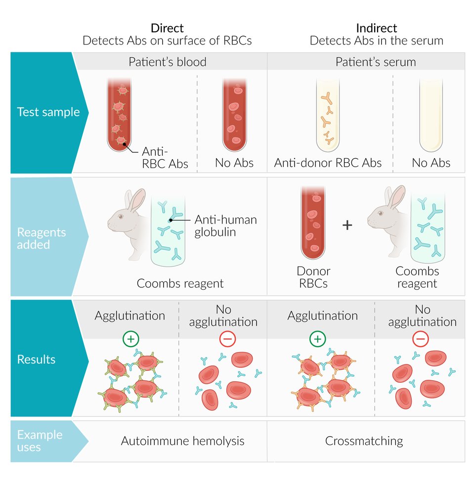
- Indirect Coombs test (detects free IgG anti-D in maternal serum)
Rising maternal anti-D titres (>15 IU/mL) indicate significant risk of fetal anemia and warrant intensified fetal monitoring.
Fetal Assessment
Ultrasound (first-line imaging):
- Hydrops fetalis signs (ascites, pleural/pericardial effusion, skin oedema, placental thickening)
- Hepatosplenomegaly, cardiomegaly
- Polyhydramnios
Middle cerebral artery (MCA) Doppler peak systolic velocity (gold standard for fetal anemia assessment):
- Normal: <1.29 MoM (multiples of median)
- Elevated PSV: >1.5 MoM indicates moderate/severe fetal anaemia and predictor of need for intrauterine transfusion
- Replaces amniotic fluid bilirubin analysis as the non-invasive standard
| Important - Question Idea | |
| The Indirect Coombs test (screening) detects FREE IgG anti-D in maternal serum and guides the risk of fetal disease. The Direct Coombs test (confirmatory) on neonatal blood detects ANTIBODY-BOUND erythrocytes in the neonate and confirms HDFN. Do NOT confuse them: indirect = maternal blood (predicts risk), direct = newborn blood (confirms haemolysis). Both may be positive in the same case, but they answer different clinical questions. | تذكر |
Neonatal Diagnosis
Direct Coombs (Antiglobulin) test (confirmatory): Strongly positive in Rh incompatibility — confirms antibody coating of neonatal erythrocytes.
Full blood count:
- Anemia (may be profound)
- Reticulocytotic (bone marrow response)
- Spherocytes and nucleated RBCs on blood film
Serum bilirubin (unconjugated): Elevated, often rising rapidly in first 24–48 hours. Level guides phototherapy vs. exchange transfusion threshold.
MANAGEMENT
Prenatal Management
Monitoring:
- Serial maternal anti-D antibody titres (baseline, then every 2–4 weeks after 24 weeks)
- MCA Doppler assessment (every 1–2 weeks once titres >15 IU/mL or if PSV elevated)
- Detailed ultrasound for hydrops signs
Intrauterine transfusion (IUT) — indicated if fetal anemia severe (MCA PSV >1.5 MoM, positive cordocentesis, or hydrops present):
- Route: Umbilical vein (first-line), intrahepatic vein, or intraperitoneal (less common)
- Blood: O-negative, screened, irradiated, crossmatched
- Repeat transfusions every 2–4 weeks until fetal maturity or delivery
IV immunoglobulin (IVIG): 2 g/kg weekly in selected severe cases (adjunct therapy, evidence mixed); may reduce hemolysis rate.
Early delivery: Consider at 37+ weeks if severe disease; must balance fetal maturity against ongoing in-utero hemolysis.
Postnatal Management
Phototherapy: Start at gestation-dependent thresholds (lower thresholds in prematurity, Rh disease). Intensive phototherapy for rising unconjugated bilirubin.
Exchange transfusion: Performed if unconjugated bilirubin approaches neurotoxic threshold (gestation-dependent) despite phototherapy. Uses O-negative blood with same ABO/RhD as infant.
Supportive RBC transfusion: For significant anemia (Hb <100 g/L in premature infant, <130 g/L in term infant presenting within 48 hours).
IVIG: 0.5–1 g/kg IV may reduce hemolysis and need for exchange transfusion (use in addition to phototherapy).
Supportive care:
- Monitor for hypoglycaemia (from liver dysfunction and metabolic demand)
- Correct hypocalcaemia (from haemolysis and liver dysfunction)
- Manage heart failure and pulmonary oedema if hydrops present
- Iron supplementation post-discharge (compensatory erythropoiesis)
PREVENTION & ANTI-D PROPHYLAXIS
Prevention of RhD alloimmunisation via anti-D immunoglobulin is highly effective and remains the cornerstone of management. The goal is to prevent maternal sensitization before it occurs.
Anti-D Immunoglobulin (RhIG) Prophylaxis Regimens
Routine antenatal prophylaxis (for all unsensitized RhD-negative women):
- 500 IU (100 μg) IM at 28 weeks and 34 weeks, OR
- 1500 IU (300 μg) single IM dose at 28 weeks (alternative, country-dependent)
Following sensitizing events (within 72 hours is critical):
- Dose calculated from fetomaternal hemorrhage (FMH) testing
- Standard: 500 IU covers ~4 mL fetal whole blood (2 mL fetal RBCs)
- Additional dosing if FMH exceeds this threshold
- Events: delivery, miscarriage, abortion, antepartum hemorrhage, trauma, invasive procedures
Postpartum prophylaxis: 500 IU IM within 72 hours of delivery if newborn is RhD-positive or RhD unknown.
| ملاحظة سريرية – Clinical Note | |
| The 72-hour window is absolute. Anti-D must be given within 72 hours of a sensitising event (including delivery) to prevent maternal IgG sensitisation. If given later, it offers no protection and a sensitised pregnancy (with IgG anti-D) may already be underway. This timing is repeatedly tested in clinical exams and is critical to clinical safety. | ملاحظة |
Fetomaternal Haemorrhage (FMH) Testing
Purpose: Quantify fetal RBCs in maternal circulation to calculate additional anti-D dose in cases of large bleeds.
Methods:
- Kleihauer-Betke (KB) test: Acid elution detects fetal Hb F (resistant to acid) vs. maternal Hb A. Semi-quantitative; observer-dependent.
- Flow cytometry: More accurate, quantifies fetal cells directly. Preferred where available.
Protocol:
- FMH test performed at delivery (postpartum blood film) and after any sensitising event
- Result >4 mL fetal RBCs (8 mL whole blood) requires additional anti-D dosing (1 unit per 2 mL fetal RBCs beyond the baseline 500 IU)
KEY POINTS FOR EXAMS
- Primary response = IgM (does NOT cross placenta): First RhD-incompatible pregnancy is usually spared. Second and subsequent pregnancies are at risk when rapid IgG response occurs.
- Sensitising events are the trigger: Delivery, miscarriage, abortion, antepartum haemorrhage, trauma, and invasive procedures all risk maternal sensitisation. Anti-D must be given within 72 hours.
- MCA Doppler peak systolic velocity is the non-invasive gold standard for assessing fetal anaemia severity. Elevated PSV (>1.5 MoM) guides the decision for intrauterine transfusion.
- Indirect Coombs (maternal serum) vs. Direct Coombs (neonatal blood): Indirect predicts risk; Direct confirms haemolysis. Both may be positive in the same case but serve different diagnostic purposes.
- ABO incompatibility can affect the FIRST pregnancy (unlike Rh, which spares it) because mothers naturally carry IgG anti-A/B from infection/vaccination. This is a common exam trap.
- Kell alloimmunisation is the second-most severe HDFN cause after RhD. It uniquely suppresses erythropoiesis (not just haemolysis), making fetal anaemia worse and transfusion-resistant.
- Anti-D prophylaxis is preventive, not therapeutic. Once a woman is sensitised (IgG anti-D detected), anti-D cannot reverse the sensitisation. Prevention is always better and is achieved with routine antenatal anti-D (28 and 34 weeks) and post-sensitising-event anti-D (within 72 hours).
- FMH testing (Kleihauer-Betke or flow cytometry) quantifies fetal RBCs in maternal blood. It guides additional anti-D dosing beyond the baseline 500 IU if FMH is large.
- Intrauterine transfusion (IUT) is indicated when MCA PSV >1.5 MoM or hydrops is evident. Routes include umbilical vein (first-line), intrahepatic, or intraperitoneal.
- Exchange transfusion remains the definitive treatment for severe unconjugated hyperbilirubinemia approaching neurotoxic thresholds despite intensive phototherapy. Threshold is gestation-dependent.
| ABO Incompatibility vs. Rh Incompatibility | ||
|---|---|---|
| Feature | ABO Incompatibility | Rh Incompatibility |
| Incidence | Common (~20% pregnancies) | Rare (post-prophylaxis) |
| First pregnancy affected | Yes, possible | Rare |
| Severity | Mild, often asymptomatic | Moderate to severe |
| Coombs test | Weak positive/negative | Strongly positive |
| Clinical course | Jaundice, mild anaemia | Severe anaemia, hydrops, kernicterus |
احصل على التجربة الكاملة
اشترك للوصول لفيديوهات الشرح التفصيلي والبطاقات التعليمية التفاعلية وأسئلة الممارسة مع تتبع التقدم.