شرح المدرسين

Summary
Recurrent pregnancy loss (RPL) is a complex, multifactorial condition affecting 1–2% of women of reproductive age. Despite significant advances in understanding its etiologies—genetic, anatomical, endocrine, immunological, and thrombotic—over half of cases remain unexplained. The emotional and psychological burden on couples is substantial, requiring compassionate multidisciplinary support. Comprehensive evaluation and targeted management of identified causes are essential. Importantly, with appropriate treatment and counselling, 50–70% of women with RPL achieve a successful live birth in the next pregnancy.
Definition & Epidemiology
Recurrent pregnancy loss (RPL), also referred to as recurrent miscarriage or habitual abortion, is classically defined as three or more consecutive pregnancy losses before 20–24 weeks of gestation. The Royal College of Obstetricians and Gynaecologists (RCOG) defines it as three or more miscarriages before 24 weeks, although in clinical practice, some guidelines recommend evaluation after two losses if there are concerning features (e.g., advanced maternal age, prior investigation results).
RPL affects approximately 1–2% of women of reproductive age, though the prevalence may be higher if including two-loss cohorts. The incidence rises with maternal age and prior miscarriage number. RPL carries significant psychological and emotional burden, requiring sensitive counselling, multidisciplinary support, and realistic prognostication. Many couples experience depression, anxiety, and relationship strain; integrated mental-health support is essential to holistic care.
Etiology & Risk Factors
RPL is multifactorial, with an identifiable cause in only 40–50% of cases. More than half of affected couples receive no clear explanation for their losses, a sobering reality that frames the need for supportive and reassuring counselling.
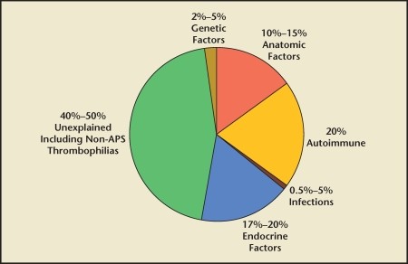
| RPL Etiologies – GAITER Mnemonic | |
|
G – Genetic (embryonic trisomy, parental translocations) A – Anatomical (septate uterus, Asherman's, cervical incompetence) I – Immunologic (Antiphospholipid Syndrome) T – Thrombophilic (Factor V Leiden, prothrombin mutation, protein C/S deficiency) E – Endocrine (diabetes, thyroid disease, PCOS, progesterone deficiency) R – Risk factors (maternal age >40, BMI, smoking, alcohol) |
جملة تذكرية |
1. Genetic Factors
- Embryonic chromosomal abnormalities account for 30–57% of miscarriages (mainly trisomies 16, 22, 21, 13). Maternal age is a strong risk factor; risk rises from 11% at age 20 to >50% after 40 and >90% above 45.
- Parental chromosomal rearrangements are present in 2–5% of RPL couples, usually balanced reciprocal or Robertsonian translocations. These lead to unbalanced conceptions with 50% risk of aneuploidy per pregnancy.
| Important – Genetic Translocation: Exam Focus | |
|
Exam Pearl: A balanced parental translocation does not cause symptoms in the carrier (clinically normal and healthy), but the risk is only unbalanced gametes. The offspring may be normal, a balanced carrier, or unbalanced (miscarriage). Genetic counselling and prenatal diagnosis (CVS/amnio) are necessary before conception. |
تذكر |
2. Anatomical Abnormalities
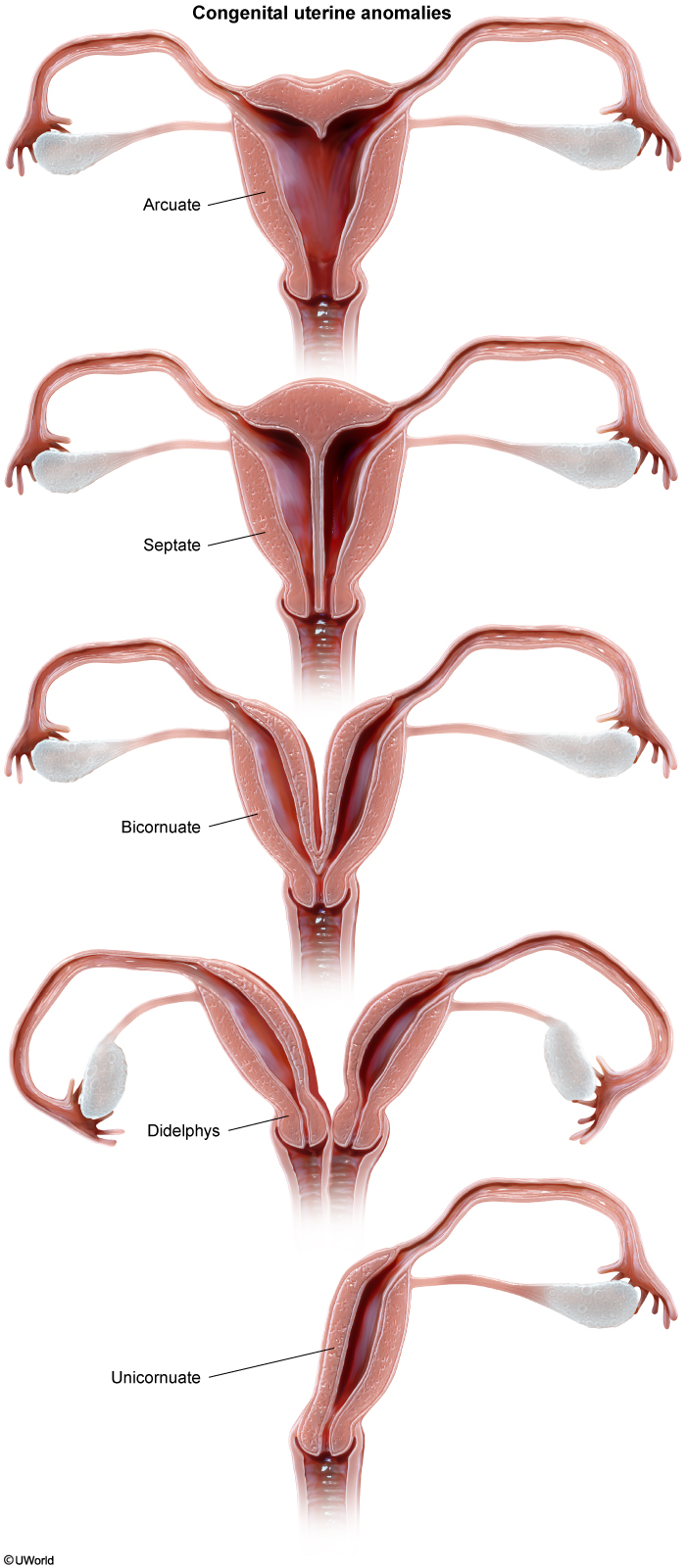
Congenital uterine malformations (resulting from Müllerian duct abnormalities) account for ~12% of RPL cases. The three most clinically tested anomalies are:
- Septate uterus (most common cause of uterine anomaly–related RPL) — a longitudinal septum divides the cavity due to incomplete involution. The septum is poorly vascularized, predisposing to early loss. Hysteroscopic metroplasty improves live birth rates by 10–15%.
- Bicornuate uterus — two separate uterine horns from incomplete lateral fusion. Associated with preterm labour and intrauterine growth restriction; limited evidence for surgical benefit.
- Arcuate uterus — a mild fundal indentation; minimal impact on fertility; rarely requires intervention.
Acquired anatomical lesions include intrauterine adhesions (Asherman's syndrome) from dilation and curettage or uterine instrumentation, which can cause mid-trimester loss due to reduced endometrial receptivity, and submucosal fibroids distorting the cavity. Diagnostic hysteroscopy with lysis of adhesions and fibroid resection is therapeutic.
| Important – Cervical Incompetence Presentation | |
|
Clinical Pearl: Cervical incompetence is the leading anatomical cause of second-trimester loss. Present with painless progressive cervical dilatation (often discovered on routine cervical length screening by transvaginal ultrasound) or preterm premature rupture of membranes (pPROM). |
تذكر |
3. Antiphospholipid Syndrome (APS)
| Important – Antiphospholipid Syndrome in RPL | |
|
Exam Pearl: APS is present in up to 15% of women with RPL. Present in untreated women: live birth rates ~10%. Key diagnostic criterion: persistent antiphospholipid antibodies (lupus anticoagulant, anticardiolipin, or anti-β2 glycoprotein I) on two or more occasions ≥12 weeks apart, plus clinical features (recurrent pregnancy loss or vascular thrombosis). Exam trap: APS on exams is always linked to recurrent early losses or vascular thrombosis. Treatment: Aspirin + Heparin, and anticoagulation is not given for unexplained cases. |
تذكر |
4. Endocrine and Metabolic Disorders
- Uncontrolled diabetes mellitus and thyroid disease increase miscarriage risk significantly. Target HbA1c <6.5% pre-conception and TSH 0.5–2.5 mIU/L; optimize prior to pregnancy.
- Polycystic ovarian syndrome (PCOS) is associated with RPL, likely due to insulin resistance, elevated luteinizing hormone (LH), and hyperandrogenism. Weight loss of 5–10% body weight improves outcomes; inositol or metformin may be indicated.
- Progesterone deficiency and hyperprolactinemia remain controversial etiologies. Some clinicians measure luteal-phase progesterone or serial hCG doubling rates, though evidence for progesterone supplementation beyond luteal support is limited.
5. Inherited Thrombophilias
- Factor V Leiden (FVL) — most common heritable thrombophilia (5% of Caucasians heterozygous); homozygous FVL carries higher risk.
- Prothrombin gene mutation (G20210A) — second most common; often coinherited with FVL as a compound heterozygote, conferring higher thrombotic risk.
- Protein C or S deficiency, Antithrombin III deficiency — rarer but strongly associated with recurrent second-trimester losses due to placental thrombosis and infarction.
Note: The role of thrombophilia screening in unexplained RPL remains debated; current guidelines suggest screening in specific contexts (e.g., family history of thrombosis, prior second-trimester loss).
Maternal Risk Factors (Merged)
- Maternal age >40 — miscarriage risk exceeds 50%; age >45 carries >90% risk. Parental age also contributes; paternal age >40 associated with increased aneuploidy.
- Lifestyle factors — smoking, alcohol use (>2 units daily), high caffeine intake (>200 mg daily), psychological stress, and abnormal BMI (both <18.5 and >30) all increase loss rates.
- Number of prior losses — risk increases with successive losses; after three consecutive losses, the risk of a fourth exceeds 40% (though counselling should emphasize the ~50–70% chance of live birth in the next pregnancy).
Clinical Evaluation & Investigations
All women presenting with RPL should undergo a systematic, cause-directed evaluation. The investigation strategy is guided by the suspected etiology, maternal age, and prior loss timing (early vs. mid-trimester).
History and Examination
- Detailed medical, obstetric, and family history, including prior loss gestations, delivery records, karyotypes (if available), and screening results.
- Thorough pelvic examination and assessment for clinical stigmata of systemic disease (e.g., lupus rash, weight changes, thyroid nodules).
- Enquire about lifestyle factors: smoking, alcohol, caffeine, stress, trauma, and partner/paternal factors (age, occupational exposures).
Applying Classification Table — Diagnostic Investigations by Suspected Etiology
| Diagnostic Investigations by Suspected Etiology: First-line and confirmatory tests mapped to RPL cause | |
| GENETIC CAUSES | Chromosomal & Parental Rearrangements |
| Karyotype (parental) | Balanced reciprocal or Robertsonian translocations present in 2–5% of RPL couples. Present in 30–57% of miscarriage products (aneuploidy). |
| IMMUNOLOGIC (APS) | Antiphospholipid Antibodies |
| Lupus anticoagulant (LA) | First-line test; requires ≥2 positive results ≥12 weeks apart for diagnosis. |
| Anticardiolipin IgG/IgM | Complement panel; medium or high titre associated with thrombosis and RPL. |
| Anti-β2 glycoprotein I | Anti-β2GPI alone (without LA or aCL) may be lower risk but requires repeat testing. |
| ENDOCRINE DISORDERS | Metabolic & Hormonal Screening |
| TSH + free T4 | Uncontrolled thyroid disease increases miscarriage risk; target TSH <2.5 mIU/L in pregnancy. |
| Fasting glucose / HbA1c | Screen for pregestational diabetes; poor glycaemic control ↑ risk significantly. |
| Prolactin, luteal progesterone | Progesterone deficiency remains controversial; some measure day-21 P4 or serum hCG doubling rate. |
| ANATOMICAL ABNORMALITIES | Imaging & Structural Assessment |
| Pelvic ultrasound (TVUS) | First-line; assess uterine cavity, septum, fibroids. Septate uterus best seen on 3D US or HSG. |
| 3D ultrasound or HSG | Gold standard for congenital anomalies; 3D US preferred for septate vs. bicornuate differentiation. |
| Hysteroscopy | Direct visualisation for Asherman's syndrome, submucosal fibroids; therapeutic role if resection needed. |
| Transvaginal US (cervical length) | Cervical cerclage indication if CL <25 mm in mid-trimester; baseline screening at 16–20 weeks. |
| THROMBOPHILIAS | Heritable and Acquired Coagulopathy |
| Factor V Leiden (FVL) | Most common inherited thrombophilia; heterozygous in 5% of Caucasians, homozygous much rarer. |
| Prothrombin gene mutation (G20210A) | Second most common; often coinherited with FVL (compound heterozygote ↑ risk further). |
| Protein C, Protein S, Antithrombin III | Deficiency is rarer; associated with early second-trimester losses and placental thrombosis. |
Management Protocols
Management of RPL is cause-specific and multidisciplinary. When an etiology is identified, targeted intervention can significantly improve live birth rates. For unexplained RPL, supportive care, psychological counselling, and reassurance form the foundation of management.
| Etiology-Specific Management of RPL | |||
|---|---|---|---|
| Etiology | Management Goal | Specific Intervention | Key Monitoring |
| Genetic (parental translocation) | Optimize conception and reduce aneuploidy | Genetic counselling; consider IVF + PGT-A; gamete donation or adoption if desired | Repeat testing pre-conception; NST in pregnancy if natural conception |
| Antiphospholipid Syndrome (APS) | Prevent thrombosis and improve placentation | Aspirin 75–150 mg daily + unfractionated heparin (UFH) or LMWH throughout pregnancy | Target therapeutic heparin level (aPTT or anti-Xa); monitor for thrombosis and bleeding |
| Endocrine (uncontrolled diabetes, thyroid) | Optimize hormonal control pre-conception | Optimize glycaemic control (HbA1c <6.5%); TSH 0.5–2.5 mIU/L; consider levothyroxine for subclinical hypothyroidism | Repeat HbA1c, TSH at 4–6 weeks; monitor in each trimester |
| PCOS | Reduce insulin resistance; optimize ovulation | Weight loss (5–10% body weight if BMI >25); inositol or metformin if indicated; counsel on lifestyle | Monitor for gestational diabetes; repeat TSH and glucose tolerance testing |
| Anatomical (septate uterus) | Restore normal uterine cavity | Hysteroscopic septum resection (metroplasty) prior to conception; improves live birth rate by ~10–15% | Assess cavity restoration on post-op US/HSG; allow 1–2 cycles healing before TTC |
| Anatomical (Asherman's) | Restore endometrial integrity | Hysteroscopic lysis of adhesions; consider oestrogen supplementation post-op to promote re-epithelialisation | Repeat hysteroscopy if recurrent symptoms; baseline sonohysterography (SHG) at follow-up |
| Cervical incompetence | Prevent painless dilation and PTL | Cervical cerclage (McDonald or Shirodkar) at 12–14 weeks if CL <25 mm or prior mid-trimester loss with CL <30 mm; consider vaginal progesterone 200–400 mg daily as adjunct | Cervical length US every 1–2 weeks from 16 weeks; monitor for labour signs; remove cerclage at 36–37 weeks |
| Inherited thrombophilia (no prior thrombosis) | Prevent placental microthrombi in pregnancy | Aspirin 75–150 mg daily or LMWH if high-risk phenotype (homozygous FVL, compound heterozygote); counsel on activity restrictions | Teach signs of DVT/PE; baseline and serial D-dimer (if available); clinical vigilance |
| Unexplained RPL (>50% of cases) | Supportive care and psychological support | Expectant management with close monitoring; consider empirical aspirin (controversial); multidisciplinary counselling | Regular check-ins; refer for counselling; offer support groups; reassure of ~50–70% live birth rate with next pregnancy |
Cervical Cerclage — Technical Notes
Indications: Documented cervical incompetence (transabdominal or transvaginal cervical length <25 mm at 16–24 weeks) or a prior mid-trimester loss with sudden painless dilatation or vaginal bleeding in the absence of infection or uterine contractions.
Timing: Place at 12–14 weeks of gestation. McDonald technique (encircles the cervix submucosal, higher in the cervical stroma, faster, reversible) is most common. Shirodkar technique (a purse-string suture placed higher, more invasive, lower removal risk) is reserved for recurrent cerclage failure.
Adjunctive therapy: Vaginal progesterone (micronized progesterone 200 mg or 17-alpha-hydroxyprogesterone caproate 250 mg) or pessary placement may improve outcomes further, especially if cervical length 20–25 mm.
Cerclage removal: Remove at 36–37 weeks to permit vaginal delivery. Elective cerclage removal before term labour avoids cervical trauma and uterine rupture from unplanned labouring with cerclage in situ.
Unexplained RPL & Prognosis
Unexplained RPL
More than 50% of RPL cases have no identifiable cause despite comprehensive investigation. This absence of explanation presents both a clinical and psychological challenge. It is crucial to reframe this uncertainty positively: unexplained RPL does NOT mean a worse prognosis—it simply reflects the limitations of current diagnostic testing.
Management approach:
- Supportive care and counselling — Detailed discussion of the ~50–70% live birth rate with the next pregnancy, regardless of treatment. Many couples benefit from the reassurance that even without intervention, over half will achieve success.
- Controversial interventions — Empirical low-dose aspirin (75–150 mg daily from pre-conception) is sometimes offered, though evidence remains mixed and conflicting. Empirical anticoagulation or immunosuppression is not recommended outside of proven etiologies (e.g., APS, thrombophilia).
- Psychological support — Referral to counselling, support groups (many nations have RPL networks), couples' therapy, and discussion of alternative family-building options (adoption, donor gametes) if losses continue.
- Lifestyle optimisation — Smoking cessation, reduced caffeine and alcohol, weight management, stress reduction, regular exercise, and folic acid supplementation (5 mg daily) are universally recommended.
Prognosis
Even without treatment, the prognosis for women with RPL is surprisingly optimistic:
- Untreated cohorts show live birth rates of 50–70% with the next spontaneous pregnancy, suggesting that many RPL patients will achieve success without intervention.
- Prognosis depends on:
- Maternal age — Age >40 carries higher aneuploidy risk and lower live birth rates; age >45 dramatically worsens outlook.
- Number and timing of prior losses — After three consecutive losses, the risk of a fourth exceeds 40%, but the cumulative live birth rate remains ~60% with supportive management.
- Underlying etiology — Causes such as APS, anatomical anomalies, and endocrine disorders have specific treatments that markedly improve outcomes. Genetic translocations have more guarded prognosis (10–20% natural conception live birth rate) but excellent outcomes with IVF + PGT-A.
- With treatment — Women with APS treated with aspirin and heparin achieve 50–80% live birth rates (up from ~10% untreated). Surgical correction of septate uterus improves rates by 10–15%. Cervical cerclage reduces preterm birth and improves viability in women with documented incompetence.
- Counselling message — Frame RPL as a challenge that, in the majority of cases, will ultimately resolve with time, supportive management, psychological care, and identification and treatment of any modifiable risk factors. The emotional resilience of couples who continue trying after RPL is remarkable, and outcomes often justify their persistence.
Key Points for Exams – نقاط مهمة للامتحانات
High-Yield Summary for Board Exams
- Definition: RPL = ≥3 consecutive pregnancy losses before 20–24 weeks. Affects 1–2% of reproductive-age women; >50% remain unexplained.
- GAITER mnemonic: Genetic, Anatomical, Immunologic (APS), Thrombophilic, Endocrine, Risk factors. Use to recall the major etiologies systematically.
- Antiphospholipid Syndrome (15% of RPL):
- Diagnostic criteria: Persistent antiphospholipid antibodies (LA, aCL, or anti-β2GPI) ≥12 weeks apart PLUS clinical features (recurrent loss or thrombosis).
- Untreated live birth rate ~10%; treated (aspirin + heparin) ~50–80%.
- Do NOT give anticoagulation for unexplained RPL without proven APS.
- Septate uterus (most common anomaly-related RPL): Hysteroscopic metroplasty pre-conception improves live birth by 10–15%. Can be differentiated from bicornuate on 3D ultrasound or MRI.
- Cervical incompetence (second-trimester pathognomonic finding): Painless cervical dilatation at 16–24 weeks. Diagnosed by cervical length <25 mm on transvaginal US. Managed with cerclage at 12–14 weeks ± vaginal progesterone. Removes cerclage at 36–37 weeks.
- Maternal age impact: Miscarriage risk rises from 11% at age 20 to >50% after age 40, and >90% above 45 (mostly aneuploidy).
- Thrombophilias: Factor V Leiden and prothrombin G20210A are most common. Second-trimester losses more common with heritable thrombophilia due to placental infarction. Screening indicated if prior thrombosis or strong family history.
- Parental chromosomal rearrangement (2–5% of RPL): Balanced translocation carrier is clinically normal but has 50% risk of aneuploidy per pregnancy. Offer genetic counselling and consider PGT-A with IVF.
- Endocrine optimisation pre-conception: TSH <2.5 mIU/L, HbA1c <6.5%, consider levothyroxine for subclinical hypothyroidism in RPL context.
- Unexplained RPL (~50%): Prognosis is NOT grim—live birth rate 50–70% with next pregnancy even without treatment. Emphasise supportive care, psychological counselling, and lifestyle modification over empirical medications.
- Investigation sequence: Start with karyotype (parental), pelvic ultrasound, antiphospholipid antibodies. Escalate imaging (3D US, HSG, hysteroscopy) if anatomical suspicion. Thrombophilia screen reserved for high-risk phenotype (family history, prior thrombosis).
- Aspirin + heparin only for proven APS or high-risk thrombophilia in pregnancy; NOT empirical therapy for unexplained RPL.
| Exam Trap – Empirical Anticoagulation | |
|
WRONG: "Give all RPL patients prophylactic heparin or aspirin to prevent thrombosis." CORRECT: Anticoagulation is ONLY indicated for proven antiphospholipid syndrome or documented inherited thrombophilia. Empirical anticoagulation in unexplained RPL wastes resources, risks bleeding, and has no benefit. The mantra is: treat the cause, not the symptom. Exam trap: Examiners love to test your knowledge on the difference between APS + thrombophilia (which warrant heparin/aspirin) versus unexplained RPL (supportive care only). If there is no confirmed diagnosis, you do not prescribe empirical anticoagulation. |
تذكر |
احصل على التجربة الكاملة
اشترك للوصول لفيديوهات الشرح التفصيلي والبطاقات التعليمية التفاعلية وأسئلة الممارسة مع تتبع التقدم.