Summary
Small bowel obstruction (SBO) is a partial or complete blockage of flow through the small intestine and one of the most common surgical emergencies. Postoperative adhesions cause most cases.
Obstruction produces proximal bowel dilation, fluid sequestration, hypovolemia and electrolyte loss; progressive distention then drives venous congestion, arterial compromise, ischemia, bacterial translocation and finally perforation.
Patients present with the classic tetrad of colicky abdominal pain, vomiting, distention and obstipation. CT abdomen with IV contrast is the diagnostic test of choice. Stable adhesive SBO is managed with NPO, nasogastric decompression and IV fluids. Peritonitis, strangulation, perforation, closed-loop obstruction or failed nonoperative therapy mandate urgent surgery.
Introduction & Etiology
Definition & burden
SBO is a partial or complete interruption of the normal flow of intestinal contents through the small intestine due to a mechanical blockage (functional impairment produces ileus, covered separately).
It is one of the most common surgical emergencies, accounting for roughly 15–20% of admissions for acute abdominal pain, about 80% of all mechanical intestinal obstructions, and more than 300,000 hospital admissions annually in developed countries.
Etiology
- Postoperative adhesions — 60–75%: the single most common cause overall.
- Hernias — 10–20%: the most common cause in a patient with no prior abdominal surgery.
- Malignancy — 5–15%.
Geography shifts the balance: in developed countries adhesions predominate, whereas in developing countries hernias remain a major cause. When a hernia is the culprit, CT shows the transition point at the hernia orifice rather than within the peritoneal cavity.
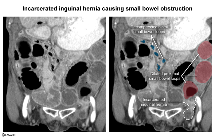
| Note | |
|
Any patient presenting with SBO who has no prior abdominal surgeries, the first thing you should do is rule out hernias, especially on groin examination. |
ملاحظة |
Pathophysiology & Classification
Classification
By mechanism:
- Mechanical SBO (adhesions, hernias, tumors, strictures)
- Functional obstruction / ileus (postoperative ileus, electrolyte abnormalities, sepsis, medications)
By degree:
- Partial — some intestinal contents still pass, often managed conservatively
- Complete — no passage of gas or stool, with a greater likelihood of surgery
By vascular status:
- Simple (no vascular compromise)
- Strangulated (compromised blood supply)
- Closed-loop obstruction, in which two points of obstruction isolate a bowel segment — carrying the highest risk for ischemia, necrosis and perforation
Pathophysiology — a staged cascade
Once flow is blocked, SBO progresses through a predictable five-stage cascade that ends in perforation if untreated.
| Pathophysiology of SBO — the 5 stages | |
|---|---|
| Stage 1 — Obstruction | |
| Mechanism | Consequence |
| Mechanical block halts passage of gas, fluid and luminal contents | Proximal bowel dilates; distal bowel collapses |
| Stage 2 — Fluid sequestration | |
| Mechanism | Consequence |
| Up to 6–8 L/day of saliva, gastric, biliary, pancreatic and intestinal secretions pool in the lumen | Hypovolemia, dehydration, hypokalemia, hypochloremia |
| Stage 3 — Rising intraluminal pressure | |
| Mechanism | Consequence |
| Distention obstructs venous outflow first, then arterial inflow | Bowel-wall edema and congestion → ischemia and infarction |
| Stage 4 — Bacterial translocation | |
| Mechanism | Consequence |
| Damaged mucosa allows bacteria and toxins to cross the bowel wall | Sepsis and septic shock |
| Stage 5 — Perforation | |
| Mechanism | Consequence |
| Persistent ischemia progresses unchecked | Necrosis, gangrene, perforation and diffuse peritonitis |
Clinical Presentation
Classic tetrad
SBO classically presents with four cardinal features: colicky, intermittent, periumbilical abdominal pain that becomes constant and severe once strangulation develops; vomiting; abdominal distention; and obstipation — failure to pass both stool and flatus, which suggests complete obstruction.
| Mnemonic – جملة تذكرية: the classic SBO tetrad | |
|
Picture a blocked drainpipe and read it top to bottom:
C-V-D-O = the four cardinal features of small bowel obstruction. |
جملة تذكرية |
The level of obstruction shapes the picture:
| Proximal vs Distal SBO — presentation | ||
|---|---|---|
| Feature | Proximal SBO | Distal SBO |
| Vomiting | Early, frequent, bilious | Late, feculent |
| Abdominal distention | Minimal | Marked |
| Fluid & electrolyte loss | Pronounced early (from early emesis) | Develops later |
Physical examination
General signs reflect volume loss: dehydration, tachycardia and orthostatic hypotension.
- Inspect for distention, surgical scars and hernias.
- On auscultation, bowel sounds are early hyperactive and high-pitched ("tinkling"), becoming decreased or silent late.
- Palpation reveals mild tenderness in simple SBO but localized tenderness, guarding and rebound once strangulation supervenes.
Red flags for strangulation — treat as bowel ischemia until proven otherwise: continuous (non-colicky) pain, fever, tachycardia, leukocytosis, metabolic acidosis and peritonitis.
Diagnosis
Laboratory studies
- CBC: leukocytosis and hemoconcentration.
- Electrolytes: hypokalemia and hypochloremia are frequent (secretion loss + vomiting).
- Serum lactate: elevation suggests ischemia / strangulation — but a normal lactate does NOT exclude ischemia.
| ملاحظة سريرية – Clinical note | |
|
A normal serum lactate does not rule out bowel ischemia — always rely on the patient's clinical picture. |
ملاحظة |
Imaging
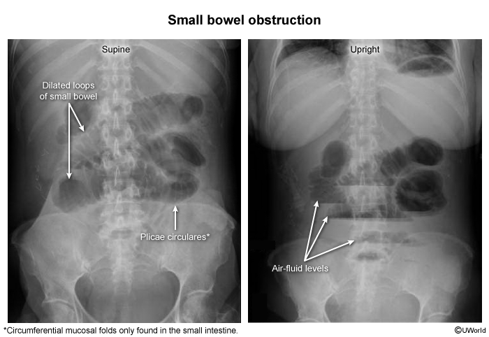
Plain abdominal radiography (first-line screen): the upright film shows multiple air-fluid levels; the supine film shows dilated loops of small bowel (diameter >3 cm) with valvulae conniventes crossing the entire lumen.
CT abdomen with IV contrast (gold standard, most sensitive): identifies the transition point, the cause, closed-loop obstruction, ischemia and perforation. SBO shows dilated proximal bowel with collapsed distal bowel; features of strangulation add bowel-wall thickening, mesenteric edema, pneumatosis intestinalis, portal venous gas and reduced bowel-wall enhancement.
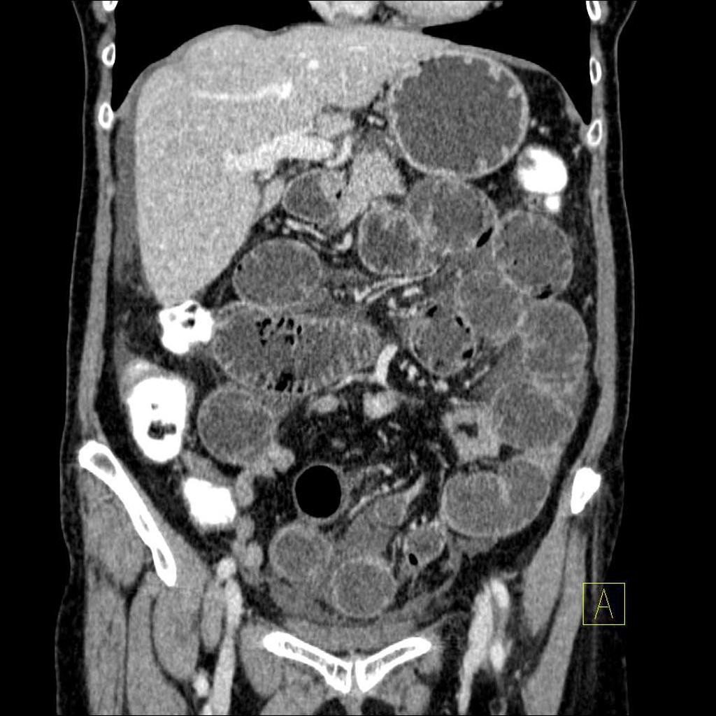
SBO must be distinguished from adynamic (paralytic) ileus, which shows gas distributed diffusely through small and large bowel with no mechanical transition point and absent bowel sounds. See the mechanical SBO versus adynamic ileus comparison for the differences in etiology, bowel sounds and radiographic pattern.
Management, Complications & Prevention
Every patient starts with resuscitation and decompression; the key decision is then nonoperative trial versus operative intervention.
| Treatment steps — from resuscitation to the operating room | |
|---|---|
| Step 1 — Initial resuscitation (ALL patients) | |
| Intervention | Detail |
| ABCs + NPO + aggressive isotonic IV fluids | Correct hypovolemia and electrolytes; place nasogastric tube for decompression and a urinary catheter to monitor output; add broad-spectrum antibiotics if strangulation, perforation or sepsis is suspected |
| Step 2 — Nonoperative management | |
| Intervention | Detail |
| NPO, NG decompression, IV fluids, serial abdominal exams | Reserved for adhesive SBO with no peritonitis, no ischemia and hemodynamic stability; succeeds in ~65–80% of cases |
| Step 3 — Operative management | |
| Intervention | Detail |
| Adhesiolysis, bowel resection, hernia repair or tumor resection | Adhesiolysis is the most common procedure; resect necrotic/perforated/non-viable bowel; repair incarcerated or strangulated hernia |
Indications for surgery
Proceed to urgent operation for: peritonitis, perforation, strangulation, closed-loop obstruction, an irreducible hernia, clinical deterioration, or failure of conservative treatment.
A complete obstruction (no gas in the rectum on imaging), especially after recent surgery, should not be observed indefinitely — delay risks ischemia.
| Exam trap | |
|
Closed-loop obstruction is considered the highest risk for ischemia and perforation, and requires urgent operative intervention. |
Note |
| Important – فكرة سؤال | |
|
Over-resuscitating an SBO patient with large volumes of 0.9% normal saline can produce a hyperchloremic non-anion-gap metabolic acidosis — a classic iatrogenic pitfall tested on boards. Use balanced crystalloids and monitor chloride. |
تذكر |
The high-yield SBO summary and treatment algorithm lays out the conservative-versus-surgical decision pathway, key imaging findings and complications at a glance.
Complications
- Early: dehydration, acute kidney injury, aspiration pneumonia, electrolyte disturbances.
- Late: bowel ischemia, necrosis, perforation, sepsis and death.
Prevention
- Adhesion prevention (most important): gentle tissue handling, meticulous hemostasis, laparoscopic approach when appropriate, and adhesion-barrier agents in selected cases.
- Hernia prevention: early diagnosis and elective repair of abdominal-wall hernias.
- Early postoperative measures: enhanced-recovery pathways, early ambulation and minimizing opioids.
Key Points for Exams – نقاط مهمة للامتحانات
- Most common cause of SBO overall → postoperative adhesions (60–75%).
- Most common cause with no prior surgery → a hernia until proven otherwise; examine the groin. In elderly women, a femoral hernia is a classic culprit and is especially prone to incarceration and strangulation.
- Most sensitive / gold-standard imaging → CT abdomen with IV contrast (shows the transition point, cause and signs of ischemia).
- Plain film clues → dilated loops >3 cm, valvulae conniventes crossing the entire lumen, and air-fluid levels on the upright film.
- Earliest bowel-sound finding → high-pitched "tinkling" sounds (later silent).
- Classic tetrad → colicky pain, vomiting, distention, obstipation (C-V-D-O).
- Most dangerous complication → strangulation with bowel ischemia; a normal lactate does not exclude it.
- Highest-risk anatomy → closed-loop obstruction (ischemia, necrosis, perforation) — needs urgent surgery.
- Absolute indications for surgery → peritonitis, ischemia/strangulation, perforation, closed-loop obstruction, failed conservative therapy. A complete SBO (no rectal air) after recent laparotomy warrants prompt operation, not indefinite observation.
- Stable adhesive SBO → NPO, NG decompression and IV fluids; succeeds in ~65–80%.
- Iatrogenic pitfall → excessive 0.9% saline resuscitation → hyperchloremic non-anion-gap metabolic acidosis.
- Neonatal SBO pearl → meconium ileus is strongly associated with cystic fibrosis.
احصل على التجربة الكاملة
اشترك للوصول لفيديوهات الشرح التفصيلي والبطاقات التعليمية التفاعلية وأسئلة الممارسة مع تتبع التقدم.