Sammary
The small intestine (4–6 m) is the primary organ for digestion, nutrient absorption, mucosal immunity, and endocrine regulation.
It functions through three specialized segments: the duodenum, which neutralizes acidic chyme and receives pancreaticobiliary secretions; the jejunum, responsible for the bulk of macronutrient and fluid absorption; and the ileum, dedicated to mucosal immune defense and the selective uptake of bile salts and vitamin B12.
Overview
The small intestine extends approximately 4–6 m from the pylorus to the ileocecal valve and is the body's principal site of digestion, nutrient and water/electrolyte absorption, GI hormone production, and mucosal immune defense.
It is divided into three segments with distinct functional roles:
| The Three Segments of the Small Intestine | ||
|---|---|---|
| Segment | Length | Primary function |
| Duodenum | ~25 cm | Acid neutralization & digestion |
| Jejunum | ~40% of jejunoileum | Major nutrient absorption |
| Ileum | ~60% of jejunoileum | Vitamin B12 & bile salt absorption |
Two location facts anchor the entire topic: Brunner glands are unique to the duodenum, while Peyer patches and vitamin B12 / bile-salt reabsorption belong to the terminal ileum.
Gross Anatomy: Duodenum, Jejunum & Ileum
Duodenum
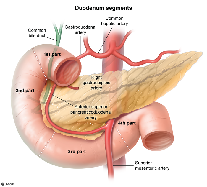
The duodenum is the shortest (~25 cm) and mostly retroperitoneal segment, forming a C-shaped loop around the head of the pancreas. It has four parts:
- First (superior) part: begins at the pylorus; contains the duodenal bulb/cap; intraperitoneal proximally.
- Second (descending) part: contains the major papilla / ampulla of Vater, which receives the common bile duct and the main pancreatic duct.
- Third (horizontal) part: crosses anterior to the inferior vena cava and aorta and passes posterior to the superior mesenteric vessels.
- Fourth (ascending) part: ends at the ligament of Treitz (duodenojejunal flexure).
The spatial relationship of the third part to the great vessels is best appreciated visually:
Jejunum vs. Ileum
The jejunum occupies the left upper abdomen and the ileum the right lower abdomen/pelvis. Their gross, vascular, and functional differences — scattered across most textbooks — are consolidated here:
| Jejunum vs. Ileum — Master Comparison | ||
|---|---|---|
| Feature | Jejunum | Ileum |
| Location | Left upper abdomen | Right lower abdomen / pelvis |
| Diameter & wall | Larger, thick wall | Smaller, thin wall |
| Color | Deep red (rich supply) | Pale |
| Plicae circulares | Tall & prominent | Sparse / absent distally |
| Arterial arcades | Few | Numerous |
| Vasa recta | Long | Short |
| Mesenteric fat | Sparse | Abundant |
| Distinctive structures | — | Peyer patches |
| Key absorption | Carbohydrate, protein, lipid | Vitamin B12 & bile salts |
| Important – فكرة سؤال | |
|
The jejunum has few arcades and long vasa recta, whereas the ileum has numerous arcades and short vasa recta. Surgeons use this to identify bowel orientation intra-operatively. |
تذكر |
Blood Supply, Venous & Lymphatic Drainage
Arterial supply tracks the embryologic foregut–midgut boundary at the ampulla of Vater:
- Proximal duodenum (foregut): superior pancreaticoduodenal artery — a branch of the gastroduodenal artery (from the celiac trunk).
- Distal duodenum (midgut): inferior pancreaticoduodenal artery — a branch of the superior mesenteric artery (SMA).
- Jejunum & ileum: SMA branches form arterial arcades, which give off vasa recta to the bowel wall.
Venous drainage: the superior mesenteric vein joins the splenic vein to form the portal vein.
Lymphatic drainage is sequential: lacteals → mesenteric nodes → superior mesenteric nodes → cisterna chyli → thoracic duct. This lacteal route is how absorbed long-chain fats (as chylomicrons) reach the systemic circulation while bypassing the portal system.
Histology of the Small Intestinal Wall
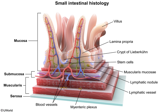
From lumen outward, the wall has four layers, each with high-yield features:
- Mucosa — simple columnar epithelium of enterocytes, goblet cells, enteroendocrine cells, and crypt-base Paneth cells; the lamina propria carries capillaries, immune cells, and lacteals; a thin muscularis mucosae drives villous movement.
- Submucosa — houses the Meissner (submucosal) plexus and, in the duodenum only, Brunner glands.
- Muscularis propria — inner circular + outer longitudinal muscle enclosing the Auerbach (myenteric) plexus, which controls peristalsis and motility.
- Serosa — visceral peritoneal covering.
| Small Bowel Epithelial & Glandular Cells | ||
|---|---|---|
| Cell type | Location | Function |
| Enterocytes | Villi | Nutrient absorption + brush-border enzymes |
| Goblet cells | Epithelium | Mucus secretion |
| Enteroendocrine cells | Epithelium | GI hormone secretion |
| Paneth cells | Crypt base | Innate immunity: lysozyme & defensins |
| Brunner glands | Submucosa (duodenum only) | Alkaline mucus → acid neutralization |
| Mnemonic – جملة تذكرية | |
|
Wall layers (lumen → outside): Mucosa → Submucosa → Muscularis propria → Serosa — “My Sister Makes Sandwiches.” Nerve plexuses: Meissner (submucosal) plexus sits in the Submucosa; Auerbach (myenteric) plexus sits between the two muscle layers of the Muscularis propria. |
جملة تذكرية |
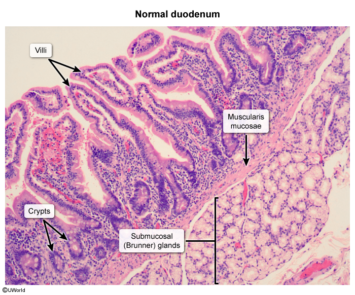
The duodenal Brunner glands are the single most useful histologic landmark distinguishing the duodenum from the rest of the small bowel:
| Important – فكرة سؤال | |
|
Brunner glands are found only in the submucosa of the duodenum and secrete alkaline mucus to neutralize gastric acid. Peyer patches are found only in the ileum. These are classic single-best-answer “location” distractors. |
تذكر |
Villi & surface-area amplification
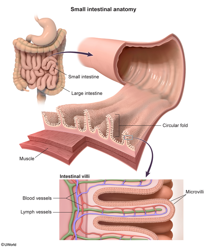
Each villus contains arterioles, venules, and capillaries — which absorb sugars and amino acids — plus a central lacteal that absorbs chylomicrons and long-chain fats. Three nested structural adaptations magnify the absorptive surface:
| Note – ملاحظة | |
|
Three structural adaptations amplify absorptive surface area in series: plicae circulares (×3) → villi (×10) → microvilli / brush border (×20), giving a combined ≈ 600-fold increase. |
ملاحظة |
Physiology: Digestion, Absorption & Motility
- Digestion: the small bowel receives pancreatic enzymes (amylase, lipase, proteases) and bile salts, which emulsify fat and form the micelles required for lipid uptake.
- Nutrient absorption is regionally specialized — the top clinically relevant sites are:
- Carbohydrates → glucose, galactose, fructose; absorbed mainly in the jejunum.
- Proteins → amino acids and di-/tripeptides; absorbed mainly in the jejunum.
- Lipids → micelles enter enterocytes, are repackaged into chylomicrons, and exit via lacteals.
- Vitamin B12 & bile salts → in the terminal ileum; B12 requires intrinsic factor, and ~95% of bile salts are reclaimed via the enterohepatic circulation.
| Note – ملاحظة | |
|
فخ امتحاني: دايماً تذكر إن الـ terminal ileum resection رح يدخل المريض بـ vitamin B12 deficiency و bile salt wasting، لأن الـ terminal ileum هو موقع الـ absorption للإثنين. الـ bile salt malabsorption بيسبّب choleretic (secretory) diarrhea. |
ملاحظة |
Motility
Three patterns dominate:
- segmentation mixes chyme and increases mucosal contact;
- peristalsis propels contents aborally;
- migrating motor complex (MMC) — the fasting "housekeeper" wave driven by motilin — sweeps residual food debris and bacteria every 90–120 minutes.
Small Bowel Endocrine Hormones
The small intestine is the largest endocrine organ, releasing peptide hormones from named enteroendocrine cells. The master summary consolidates each hormone's source cell, stimulus, and principal actions:
| Small Bowel Hormones — Master Summary | ||||
|---|---|---|---|---|
| Hormone | Location | Source cell | Stimulus | Key actions |
| Secretin | Duodenum | S cells (duodenum) | Acidic chyme | ↑ pancreatic & biliary HCO₃⁻; ↓ gastric acid |
| Cholecystokinin (CCK) | Duodenum and jejunum | I cells (duodenum/jejunum) | Fat & amino acids | Gallbladder contraction; ↑ pancreatic enzymes; relaxes sphincter of Oddi |
| GIP (Glucose-dependent Insulinotropic Peptide) | Upper small intestine | K cells (duodenum/jejunum) | Oral glucose & fat | ↑ insulin (incretin); ↓ gastric acid |
| GLP-1 | Distal ileum and colon | L cells (distal ileum/colon) | Nutrients in distal gut | ↑ insulin; ↓ glucagon; ↑ satiety |
| Peptide YY (PYY) | Ileum and colon | L cells (ileum/colon) | Fat in distal bowel | Ileal brake: ↓ motility, gastric emptying, appetite |
| Motilin | Duodenum and jejunum | M cells (duodenum/jejunum) | Fasting state | Initiates MMC every 90–120 min |
| Somatostatin | Stomach and Duodenum | D cells | Low luminal pH | Universal GI inhibitor |
| Serotonin (5-HT) | Mucosa and Myenteric Plexus | Enterochromaffin cells | Luminal stimuli | ↑ peristalsis & secretion; activates ENS |
| Mnemonic – جملة تذكرية | |
|
Enteroendocrine cell letters map to their hormones — S–I–K–L–M–D:
|
جملة تذكرية |
Clinically important extensions: GIP was the first recognised incretin; GLP-1 is the pharmacologic target of semaglutide, liraglutide, and tirzepatide; erythromycin is a motilin-receptor agonist used as a prokinetic; and somatostatin is the universal GI inhibitor (it suppresses gastrin, CCK, secretin, insulin, and glucagon). Excess serotonin from enterochromaffin-cell (carcinoid) tumors drives increased motility and secretion. See the major GI hormones — secretion sites and actions for a consolidated map that places secretin, CCK, GIP, and motilin alongside gastrin and somatostatin.
Immunologic Function & Clinical Correlations
Peyer patches are aggregated lymphoid follicles concentrated in the terminal ileum, composed of B cells, T cells, and macrophages. Overlying M cells sample luminal antigen, driving antigen surveillance, mucosal immunity, and secretory IgA production.
Because the terminal ileum performs both vitamin B12 and bile-salt reabsorption, terminal ileal resection or disease produces a predictable triad:
- Vitamin B12 deficiency — loss of the intrinsic factor–B12 uptake site.
- Bile-salt wasting — interrupted enterohepatic circulation.
- Choleretic (secretory) diarrhea — unabsorbed bile salts stimulate colonic secretion.
Two prokinetic/endocrine correlates round out the picture: motilin agonism explains why erythromycin accelerates gastric emptying, and enterochromaffin-cell carcinoid tumors illustrate the systemic effects of serotonin excess.
Key Points for Exams – نقاط مهمة للامتحانات
The consolidated, ultra-high-yield essentials for the small intestine:
| Important – فكرة سؤال | |
|
تذكر |
One structural pearl worth over-learning: jejunum = few arcades + long vasa recta; ileum = many arcades + short vasa recta.
| Note | |
|
The jejunum is responsible for most nutrient absorption, and the (terminal) ileum is responsible for vitamin B12 and bile salts. Somatostatin is a universal inhibitor, and motilin activates the MMC during fasting. |
ملاحظة |
احصل على التجربة الكاملة
اشترك للوصول لفيديوهات الشرح التفصيلي والبطاقات التعليمية التفاعلية وأسئلة الممارسة مع تتبع التقدم.