SUMMARY
Juvenile idiopathic arthritis (JIA) is the most common chronic rheumatic disease of childhood, defined by chronic joint inflammation beginning before 16 years of age, with or without extra-articular involvement. It is a clinical diagnosis of exclusion, requiring arthritis in ≥ 1 joint that persists for ≥ 6 weeks after other causes (infection, malignancy) have been ruled out.
JIA is classified into five subtypes — oligoarticular, polyarticular, systemic-onset, psoriatic, and enthesitis-related — distinguished by the number and distribution of joints involved and by their extra-articular features. Regardless of subtype, every patient develops arthritis during the disease course.
The most feared, often asymptomatic, complication is chronic anterior uveitis (highest in ANA-positive oligoarticular disease). Treatment aims to control inflammation and preserve joint function, and the overall prognosis is excellent with early diagnosis and treatment.
Epidemiology & Diagnostic Criteria
Epidemiology
JIA is the most common pediatric rheumatic disease, with arthritis as its distinguishing manifestation. Mean age of onset is 1–3 years; presentation before 6 months of age is unusual. There is an overall female predominance, with two exceptions:
- Males and females are affected equally in systemic-onset JIA (F = M).
- Males predominate in late-onset HLA-B27–related (enthesitis-related) disease.
Diagnostic criteria
Diagnosis rests on history and clinical features — there is no single confirmatory test. All of the following must be met:
- Age of onset: ≤ 16 years.
- Arthritis: in ≥ 1 joint — swelling or effusion OR limitation of motion with tenderness and increased warmth.
- Duration: persistent arthritis for ≥ 6 weeks.
- Exclusion: other causes ruled out (e.g., infection, malignancy).
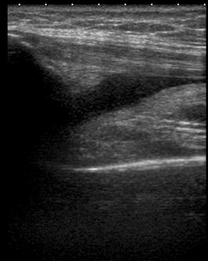
Active arthritis is defined by joint swelling or effusion; ultrasound can objectively confirm an effusion when the clinical examination is equivocal.
| Exam Trap | |
|
JIA is a diagnosis of exclusion; infection and malignancy must be ruled out first, and arthritis must persist ≥ 6 weeks before establishing the diagnosis. If the child presents with severe bone pain and pancytopenia, consider ALL rather than JIA. |
تذكر |
New diagnostic criteria for systemic JIA
Systemic-onset JIA has its own proposed criteria. Diagnosis requires fever of unknown origin — excluding infectious, neoplastic, autoimmune, and monogenic autoinflammatory diseases — documented daily (≥ 39 °C once daily, falling to ≤ 37 °C between fever spikes) for at least 3 consecutive days and recurring for at least 2 weeks, together with 2 major criteria or 1 major plus 2 minor criteria.
| New Diagnostic Criteria for Systemic JIA: Diagnosis = qualifying fever + 2 major criteria, OR 1 major + 2 minor criteria | |
| Major criteria | 2 required (or 1 if accompanied by 2 minor) |
| Evanescent erythematous rash | |
| Arthritis | |
| Minor criteria | 2 required when only 1 major is present |
| Lymphadenopathy and/or hepatomegaly and/or splenomegaly | |
| Serositis | |
| Arthralgias of ≥ 2 weeks duration | In the absence of arthritis |
| Leukocytosis (≥ 15,000/mm³) with neutrophilia | |
JIA Subtypes
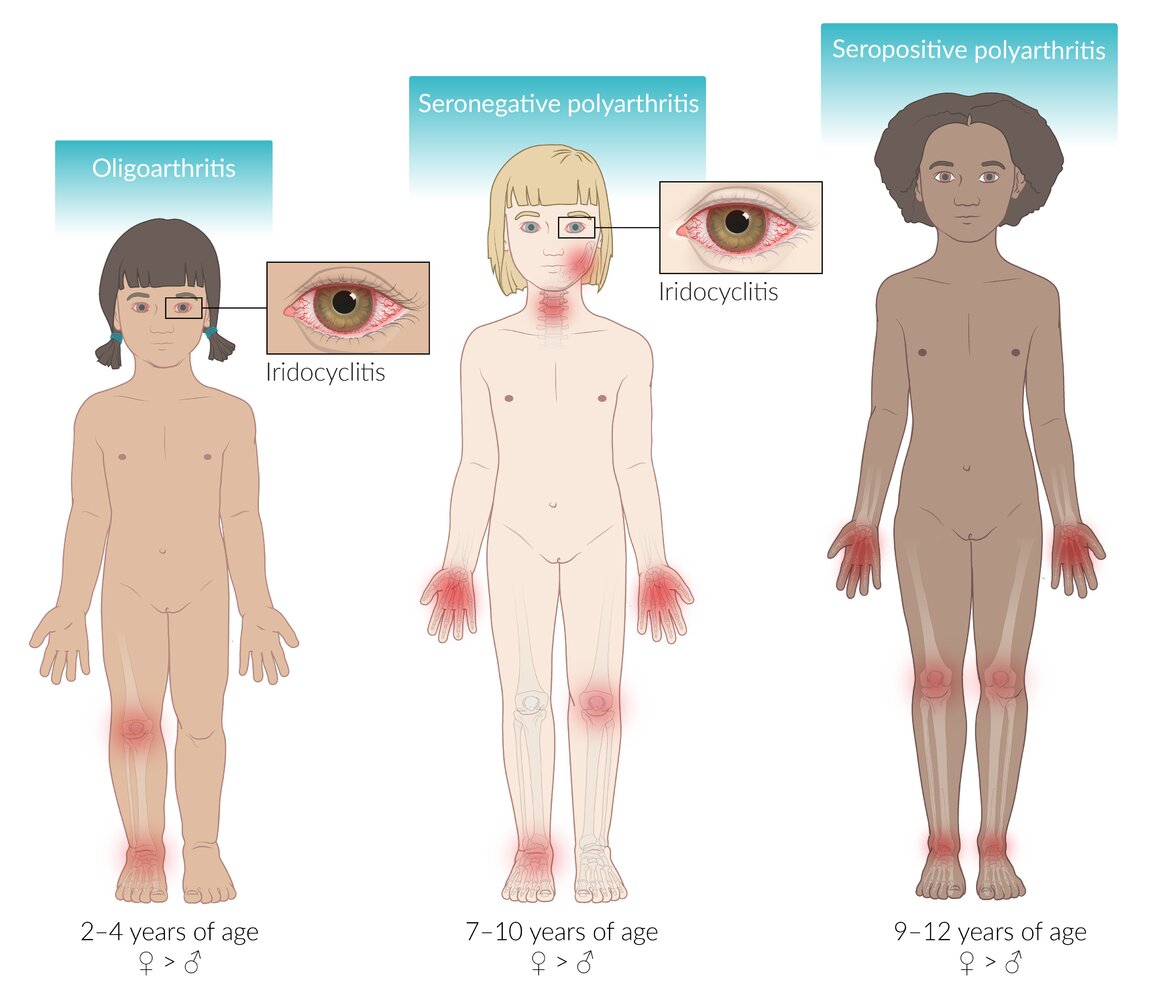
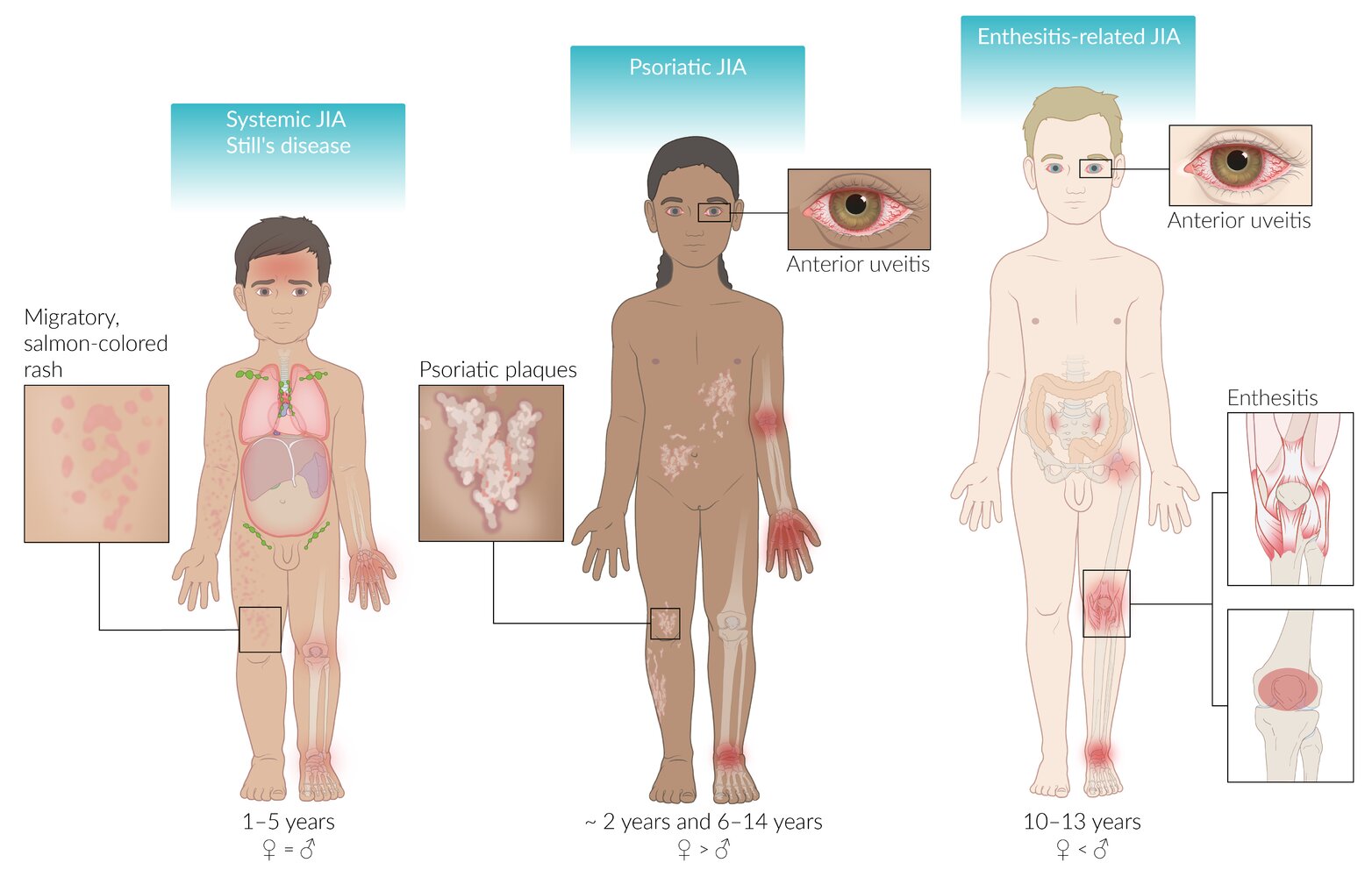
JIA is classified by the number and distribution of joints involved together with extra-articular features. The pattern of joint involvement — and its link to iridocyclitis (anterior uveitis) — varies by subtype.
| JIA Subtypes | |||||
|---|---|---|---|---|---|
| Feature | Oligoarticular | Polyarticular | Systemic-onset | Psoriatic | Enthesitis-related |
| Frequency | ~50% (most common) | 30–40% | 10–15% | 2–11% | 3–11% |
| Typical age of onset | 1–5 yr | Biphasic: 2–5 or 10–14 yr | Any age < 18 yr | Biphasic: 2–4 or 9–11 yr | Late childhood / adolescence |
| Joint pattern | < 5 joints; asymmetric; large joints, especially knees | ≥ 5 joints; symmetric; small (hands/feet) + large (knees, ankles, hips) | Arthritis in ≥ 1 joint for ≥ 6 wk; may be overshadowed by systemic features at onset | Arthritis + psoriasis, or arthritis + ≥ 2 minor criteria | Arthritis/enthesitis + sacroiliac tenderness or lumbosacral pain |
| Key associations | ANA positive in 65–85% | RF− (early & late childhood) or RF+ (> 8 yr; more severe, rheumatoid nodules) | Quotidian fever ≥ 2 wk, evanescent salmon-pink rash, hepatosplenomegaly, lymphadenopathy, serositis | Dactylitis, nail pitting, psoriasis in a first-degree relative | HLA-B27 positive; male onset > 6 yr; FHx of HLA-B27–associated disease |
| Uveitis | High risk — chronic, asymptomatic anterior uveitis | May occur | Rare | May occur | Acute, symptomatic anterior uveitis |
| Gender ratio | F > M | F > M | F = M | F > M | M > F |
Two subtypes carry additional sub-classifications: oligoarticular disease is persistent (≤ 4 joints throughout the first 6 months and beyond) or extended (≤ 4 joints initially, then > 4 joints after 6 months), while polyarticular disease is divided by rheumatoid factor status (RF− vs the more severe RF+). See the JIA subtype clinical and laboratory feature summary for a side-by-side reference of the three major subtypes.
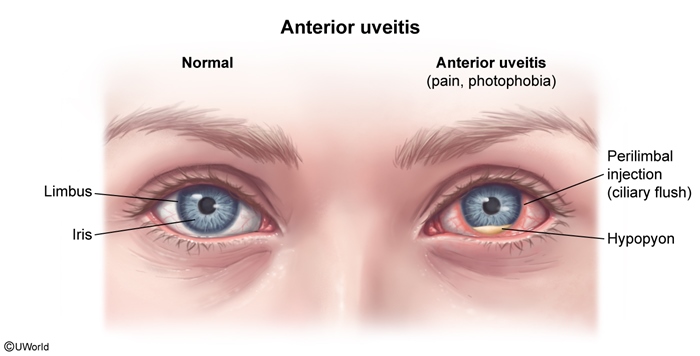
The oligoarticular subtype deserves special attention because its ANA-positive patients are at highest risk for chronic anterior uveitis — inflammation of the iris and ciliary body that is typically silent until vision is threatened.
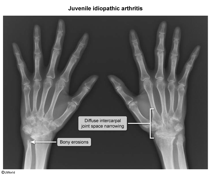
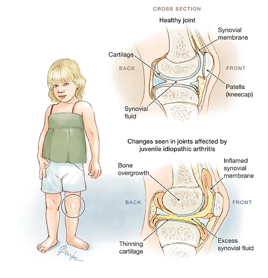
In long-standing, poorly controlled disease, chronic synovial inflammation produces radiographic joint-space narrowing and erosions.
| Clinical Note | |
|
ANA testing is ordered here to determine uveitis risk, not for diagnosis. ANA-positive oligoarticular patients carry the highest risk and require routine slit-lamp (ophthalmologic) screening even without eye pain, because uveitis in this context is chronic and asymptomatic. |
Note |
Oligoarticular JIA
- < 5 joints involved. Accounts for 50% of cases.
- Patients usually present at 1–5 years of age with a female predominance.
- A good majority (65–85%) of patients have a positive antinuclear antibody (ANA). These patients are at high risk for developing chronic uveitis (inflammation of the iris and ciliary body).
- Articular involvement may present with swelling of one or two joints, not necessarily symmetric. The most common joints involved are the knees and hips.
- Subtypes of oligoarticular JIA are recognized on the basis of the presence and timing of joint involvement:
- Persistent oligoarthritis: patients with one to four joints involved in the first 6 months of disease who do not develop arthritis in additional joints.
- Extended oligoarthritis: patients with one to four joints involved during the first 6 months of disease who then develop arthritis in greater than four joints.
Polyarticular JIA
- Five or more joints involved. Accounts for approximately 30–40% of cases, with systemic involvement and extra-articular features generally mild or absent.
- Affects females more frequently than males.
- Articular involvement is characterized by a symmetric polyarthritis. It typically involves both small joints (hands and feet) and large joints (knees, ankles, and hips).
- Subtypes of polyarticular JIA are based on the absence or presence of serum rheumatoid factor:
- RF− disease: presents both early and late in childhood.
- RF+ disease: presents in children older than 8 years. More severe than RF− disease, with a higher risk of severe arthritis and rheumatoid nodules.
Systemic-onset JIA
- Accounts for 10–15% of cases. At presentation, severe systemic symptoms may overshadow symptoms of arthritis.
- High spiking fevers (temperature > 39°C) occur most commonly in the late afternoon or evening and subsequently return quickly to baseline or subnormal levels (quotidian fever pattern).
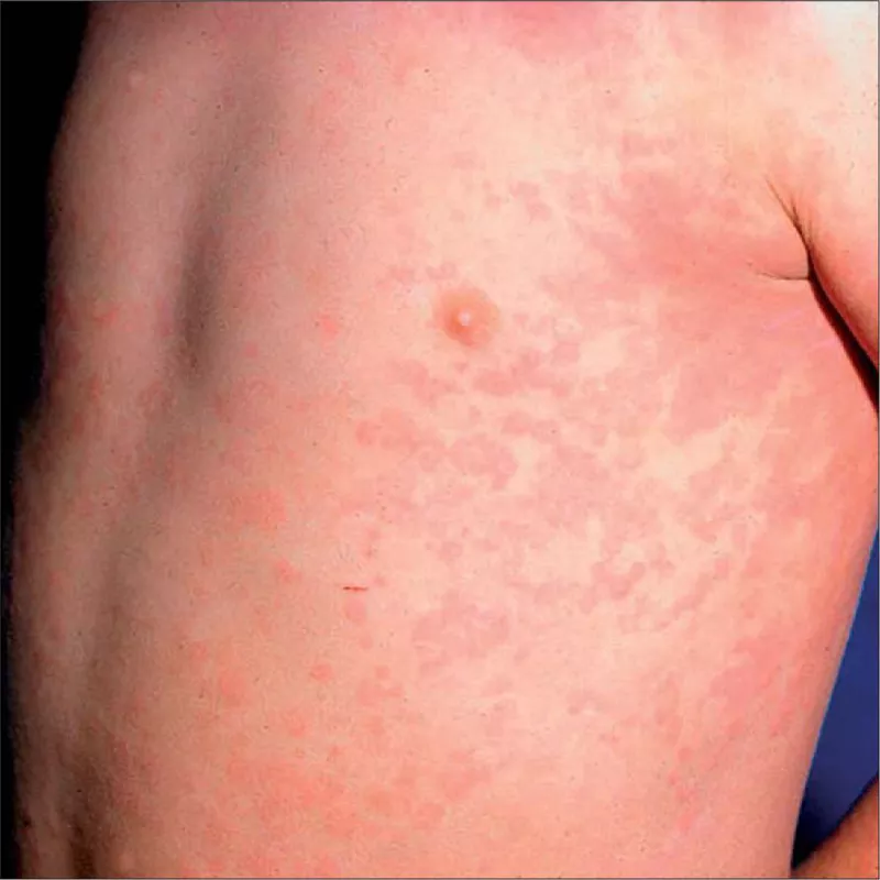
- Transient salmon-pink macular rash is most commonly found on the trunk and proximal extremities, especially during febrile episodes. The rash is evanescent (occurs with fever spikes and then fades) and nonpruritic.
- Hepatosplenomegaly.
- Lymphadenopathy.
- Other features include:
- Fatigue, anorexia, weight loss, and failure to thrive.
- Serositis, including pericarditis and pleuritis.
- CNS involvement, including meningitis and encephalopathy.
- Myositis and tenosynovitis.
SYSTEMIC-ONSET JIA HIGHLIGHTS
Systemic-onset JIA (Still disease) accounts for 10–15% of cases and is the only subtype with an equal sex distribution (F = M). At presentation, severe systemic symptoms may overshadow the arthritis.
The hallmark features are:
- Quotidian fever: high spikes > 39 °C, most commonly in the late afternoon or evening, returning quickly to baseline or subnormal levels.
- Evanescent salmon-pink macular rash: non-pruritic, on the trunk and proximal extremities, appearing with fever spikes and then fading.
- Hepatosplenomegaly.
- Lymphadenopathy.
The most common non-specific systemic complaints are fatigue, anorexia, and weight loss. Other organ involvement may include serositis (pericarditis and pleuritis) and, less commonly, CNS involvement, myositis, and tenosynovitis.
| Systemic-onset JIA (Still disease) — "FRESH" – جملة تذكرية | |
|
F — Fever: quotidian spiking fever > 39 °C, late afternoon/evening, returns to baseline R — Rash: evanescent salmon-pink macular rash, non-pruritic, flares with fever E — Enlarged organs: hepatosplenomegaly S — Swollen nodes: lymphadenopathy H — Hot/hurting joints: arthritis (± serositis) |
جملة تذكرية |
| Clinical Note | |
|
The fever here is quotidian — it spikes above 39°C once a day, usually in the afternoon or evening, and returns to baseline or subnormal levels. The salmon-pink rash appears with the fever spike and then fades (evanescent). Remember: systemic JIA is the only subtype with equal sex distribution (F = M). |
Note |
LABORATORY FINDINGS
Laboratory results are nonspecific; when present, they reflect the presence or extent of inflammation and are most useful for evaluating other disease processes.
Serum (markers of inflammation): elevated ESR and CRP, thrombocytosis (elevated platelet count), hyperferritinemia, and hypergammaglobulinemia.
Hematology: anemia that is typically microcytic and hypochromic, consistent with anemia of chronic disease.
Rheumatoid markers:
- Rheumatoid factor (RF): negative in most patients. It is not diagnostic, but RF positivity helps determine prognosis in polyarticular disease (more severe arthritis, rheumatoid nodules).
- Antinuclear antibody (ANA): present in 65–85% of oligoarticular and ~50% of polyarticular disease, but rare in systemic-onset JIA. ANA is not a diagnostic test — it stratifies the risk of uveitis.
| فخ امتحاني | |
|
RF is not diagnostic for JIA and is usually negative. But if it is positive in a polyarticular patient older than 8 years, it indicates worse prognosis: more severe arthritis + rheumatoid nodules. Note: ANA is rare in systemic-onset JIA. |
تذكر |
MANAGEMENT
The goals of treatment are to decrease joint inflammation and preserve function. Management combines pharmacologic control of inflammation with physical, surgical, and supportive measures.
- Control of inflammation: NSAIDs are first-line to reduce pain and inflammation; immunomodulatory agents — glucocorticoids, methotrexate, sulfasalazine, and TNF inhibitors — are used for more severe symptoms.
- Mechanical / physical measures: physical and occupational therapy to maintain range of motion and function.
- Surgery: joint replacement or synovectomy, generally reserved for recalcitrant joint contractures or destruction.
- Supportive: routine ophthalmologic (slit-lamp) screening for uveitis and psychosocial support.
Prognosis: the outlook is excellent with appropriate early diagnosis and treatment; the incidence of complications depends on the type and subtype of JIA. Refer to the comprehensive JIA overview (epidemiology, treatment, and complications) for a consolidated reference on management and key complications such as uveitis and bone erosion.
| Clinical Note | |
|
Start with NSAIDs for pain and inflammation; reserve immunomodulators (glucocorticoids, methotrexate, sulfasalazine, TNF inhibitors) for more severe cases. And do not forget: ophthalmologic screening for uveitis is an essential part of management, not just medication. |
Note |
KEY POINTS FOR EXAMS
| Important – نقاط مهمة للامتحانات | |
|
تذكر |
For a single-source review of how the three major subtypes differ at the bedside and in the lab, see the systemic vs polyarticular vs oligoarticular JIA feature summary.
احصل على التجربة الكاملة
اشترك للوصول لفيديوهات الشرح التفصيلي والبطاقات التعليمية التفاعلية وأسئلة الممارسة مع تتبع التقدم.