Summary
Weakness in a child is reduced ability to move muscles against resistance, localized to upper motor neuron, lower motor neuron, neuromuscular junction, or muscle.
1. Guillain-Barré syndrome (GBS) is acute, post-infectious demyelinating polyneuropathy, typically 1–4 weeks after Campylobacter jejuni gastroenteritis, presenting with ascending symmetric flaccid paralysis, areflexia, and downgoing Babinski. Diagnosis is clinical with albuminocytologic dissociation in CSF and slowed nerve conduction; treat with IVIG or plasmapheresis and respiratory monitoring; most recover completely.
2. Duchenne muscular dystrophy (DMD) is X-linked recessive dystrophin disorder: boys aged 2–3 years present with proximal weakness, Gowers sign, calf pseudohypertrophy, markedly elevated CK, confirmed by genetic testing.
3. Becker muscular dystrophy (BMD) is milder with reduced dystrophin and later onset.
Key complications: respiratory failure in GBS and cardiomyopathy in dystrophies.
Overview
Weakness means a reduced ability to purposefully move muscles against resistance. It is the presenting symptom of neuromuscular disease, so the first task is to localize it.
Four anatomical categories of weakness
- Upper motor neuron disorders: spasticity, hyperreflexia, upgoing Babinski.
- Lower motor neuron disorders (anterior horn cell, nerve root, peripheral nerve): flaccid weakness, hypo-/areflexia. GBS belongs here.
- Neuromuscular junction disorders: fatigable weakness; reflexes and sensation usually preserved.
- Myopathies: proximal weakness, no sensory symptoms, reflexes preserved until late. The muscular dystrophies belong here.
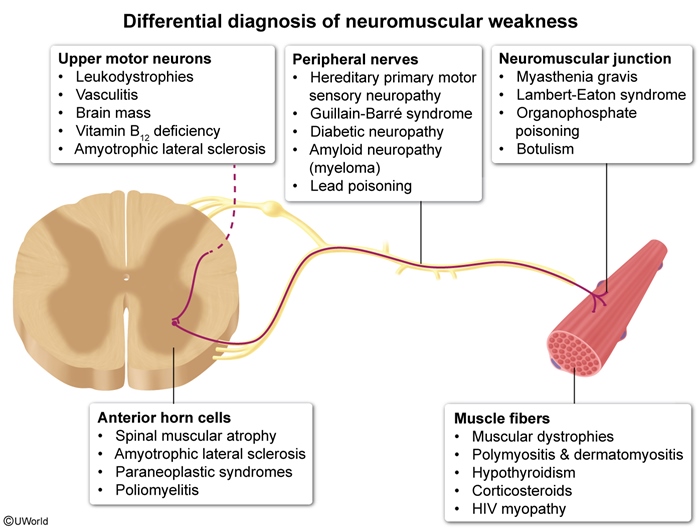
Roadmap of this lesson
- Guillain-Barré syndrome: acute, acquired peripheral nerve disease.
- Duchenne muscular dystrophy: chronic, inherited myopathy that starts in early childhood.
- Becker muscular dystrophy: the milder dystrophin disorder that starts later.
Guillain-Barré Syndrome: Etiology, Pathophysiology & Clinical Features
Definition
GBS (acute inflammatory demyelinating polyneuropathy, a demyelinating polyneuritis) is an acute peripheral nerve disease. It causes ascending weakness, areflexia and paresthesias.
Etiology
- Campylobacter jejuni: the most common associated infection, causing a prodromal gastroenteritis 1–4 weeks earlier.
- Other commonly tested triggers:
- Mycoplasma pneumoniae
- Cytomegalovirus (CMV)
- Epstein-Barr virus (EBV)
- Influenza
Pathophysiology
- Molecular mimicry: the immune response raised against the infection (activated T cells and antibodies) cross-reacts with antigens on the Schwann cell membrane.
- Main sites of demyelination: the ventral (motor) spinal roots and peripheral myelinated nerves.
- Histology shows an endoneurial infiltrate of lymphocytes and macrophages that strip the myelin, causing segmental demyelination.
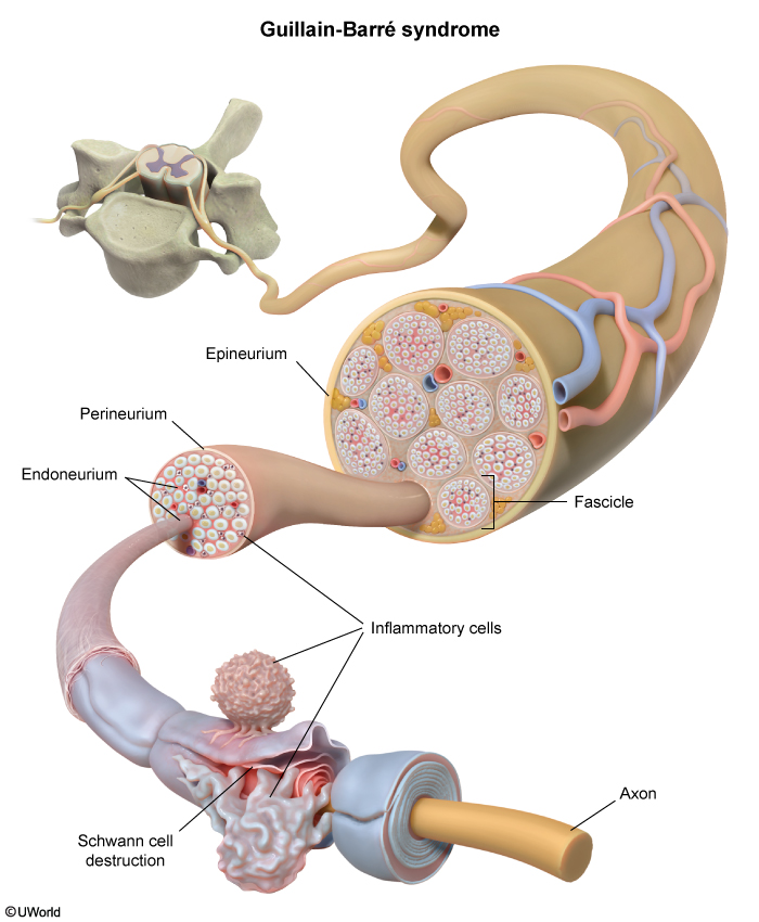
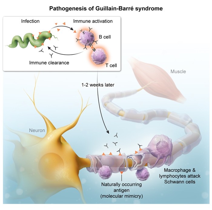
Clinical features
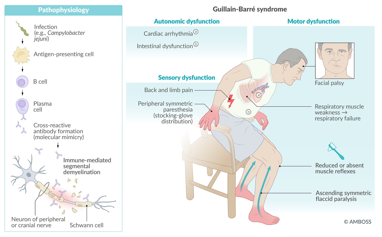
- Classic scenario: a previously well, afebrile child whose leg weakness rapidly spreads upward. The child may refuse to walk or have leg or back pain.
- Ascending, symmetric flaccid paralysis that can reach the respiratory muscles and cause respiratory arrest.
- Sensory loss is rare and limited to vibration and position sense. Distal tingling is common.
- Cranial nerves: facial weakness in 40–50%, often bilateral. Pupils are spared.
- Tendon reflexes absent. Babinski sign downgoing (normal).
- Less often: autonomic instability and ataxia.
- Bladder function is usually normal.
| فخ امتحاني | |
|
Exam trap: Even if the patient has severe ascending paralysis and areflexia, in GBS bladder function remains normal and the Babinski sign is downgoing. If you see early urinary retention or upgoing Babinski or sensory level, think of acute spinal cord compression, not GBS. |
تذكر |
| Note | |
| Miller Fisher syndrome, a variant of Guillain–Barré syndrome, is characterized by ophthalmoplegia, ataxia, and areflexia. | ملاحظة |
For a side-by-side contrast with a cord lesion, see the comparison of transverse myelitis vs. Guillain-Barré syndrome (sensory level, sphincters, MRI and CSF).
Miller Fisher syndrome (variant)
Miller Fisher syndrome is a GBS variant defined by the triad of ophthalmoplegia, ataxia and areflexia.
| Mnemonic – جملة تذكرية: Miller Fisher syndrome | |
|
“Fisher is O-A-A” — he can't look, can't walk straight, and can't jerk:
Limb strength is often relatively preserved; the variant is strongly linked to anti-GQ1b antibodies (GQ1b is concentrated in CN III, IV and VI). |
جملة تذكرية |
Guillain-Barré Syndrome: Diagnosis, Management & Prognosis
Diagnosis
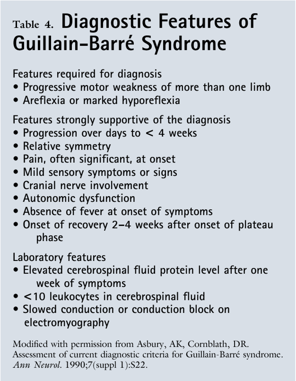
The diagnosis is clinical (acute ascending symmetric weakness with areflexia). Tests support it and rule out mimics.
- Step 1: Assess breathing first. Do bedside spirometry (forced vital capacity, negative inspiratory force) and repeat it often. An FVC <20 mL/kg, bulbar weakness or severe dysautonomia calls for ICU care and elective intubation.
- Step 2: Lumbar puncture and CSF analysis show albuminocytologic dissociation: ↑ protein with a normal cell count. It is usually evident about 1 week after symptom onset. Compare the patterns in the CSF findings in meningitis and GBS.
- Step 3: Electromyography / nerve conduction studies show decreased nerve conduction velocity or conduction block, which is evidence of demyelination.
- Step 4: Spinal MRI may be needed in children <3 years to rule out a compressive spinal cord lesion. In GBS the cord is normal, and the anterior nerve roots or cauda equina may enhance.
| ملاحظة سريرية | |
|
Clinical note: Albuminocytologic dissociation (elevated CSF protein with normal cell count) takes approximately a week from symptom onset to appear, so an early LP can come back normal. In children under 3 years we may need spinal MRI to rule out a compressive lesion. |
ملاحظة |
Management
- Start treatment as soon as the diagnosis is made because respiratory muscles can become paralysed.
- First-line immunotherapy (equally effective; choose one):
- IVIG: total 2 g/kg IV, split over 2–5 days. It is usually preferred in children because it is easier to give.
- Plasmapheresis (plasma exchange).
- Indicated in children who cannot walk or whose disease is still progressing, ideally started within the first 2 weeks.
- Supportive care: serial FVC, cardiac and blood pressure monitoring for dysautonomia, ventilation if needed.
- Glucocorticoids do not help in GBS.
| Important – فكرة سؤال: Albuminocytologic dissociation | |
|
تذكر |
The GBS summary table puts features, tests and treatment on one page.
Prognosis
- Complete recovery is the rule in children. Speed and completeness depend on how severe and widespread the weakness was.
- Physical therapy may be needed for several weeks or longer.
Duchenne Muscular Dystrophy (DMD)
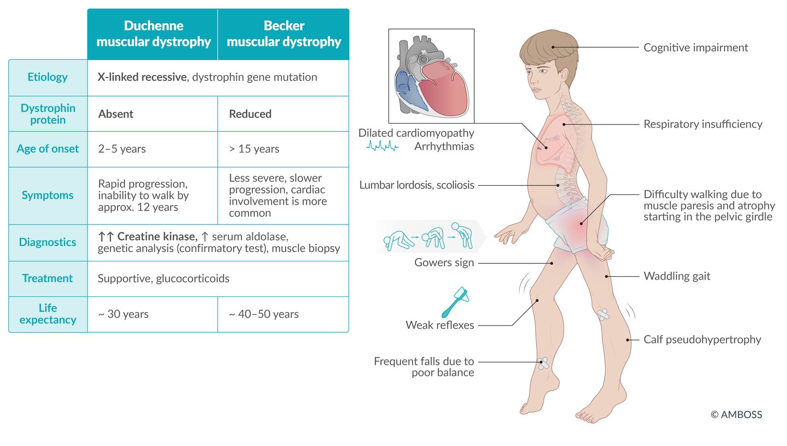
Muscular dystrophies are degenerative muscle disorders with progressive muscle wasting and replacement of skeletal muscle by fat and fibrous tissue.
Epidemiology and genetics
- Most common muscular dystrophy of childhood.
- X-linked recessive: the mother is usually a carrier, but up to about one-third of cases are new (spontaneous) mutations.
- Mutation in the dystrophin gene on Xp21 → absent or deficient dystrophin, the protein that anchors the muscle cytoskeleton to the sarcolemma. Without it, muscle fibers break down.
- Memory hook: Duchenne = Deleted dystrophin.
Clinical features
- Boys aged 2–6 years, typically 2–3 years. They often walk late and cannot keep up with their peers.
- Proximal muscle weakness: frequent falls, waddling gait, toe walking, trouble with stairs and running.
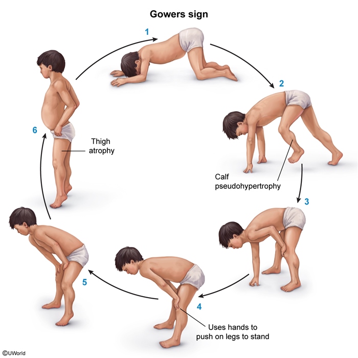
- Calf pseudohypertrophy: the calves look big because muscle has been replaced by fat and fibrous tissue.
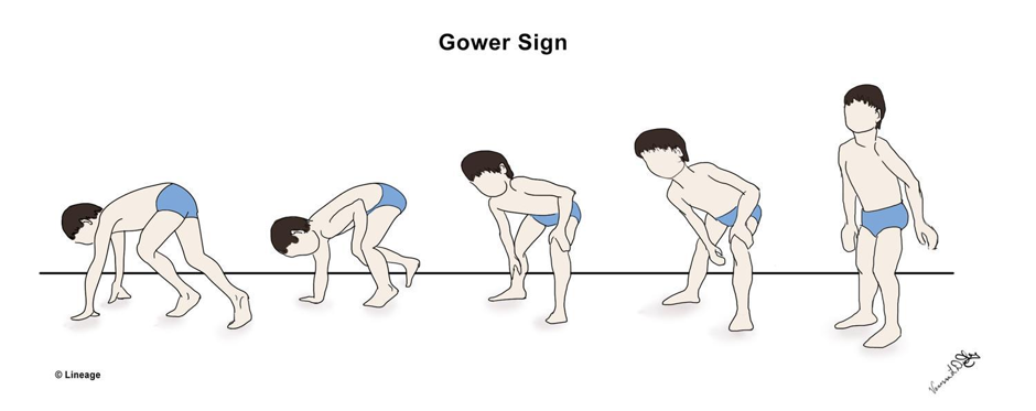
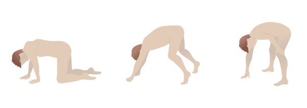
- Gowers sign: to rise from the floor, the child pushes up with the hands, climbing them up the thighs.
- Loss of ambulation by about 12 years; wheelchair-dependent by adolescence.
- Scoliosis develops after loss of walking and further restricts lung function.
- Dilated cardiomyopathy (and arrhythmias).
- Respiratory muscle weakness follows the gross motor weakness. Eventually the child cannot clear secretions, and aspiration and infection follow.
Diagnosis
- Step 1: Serum creatine kinase (CK): best initial test; markedly elevated. ALT and AST are also raised because they come from damaged muscle.
- Step 2: Genetic testing for a DMD (dystrophin) gene mutation: confirmatory test of choice.
- Step 3: Muscle biopsy: only if genetic testing is negative. It shows fibrosis, fat and degenerating muscle fibers, and absent dystrophin on immunostaining.
- At diagnosis: echocardiogram and ECG to screen for cardiomyopathy. Pulmonary function testing once the child can cooperate.
| فخ امتحاني | |
|
Testing pitfall: The elevated AST and ALT in DMD come from skeletal muscle breakdown along with the CK, not from the liver—don't pursue a liver disease workup. Confirmation is with genetic testing (gold standard), but muscle biopsy is performed only if genetic testing is negative. |
تذكر |
Management and prognosis
- Mainly supportive and multidisciplinary: physiotherapy, orthopedic, cardiac and respiratory care.
- Glucocorticoids (prednisone about 0.75 mg/kg/day or deflazacort about 0.9 mg/kg/day, orally) slow the weakness and delay wheelchair use.
- Prognosis: death at 20–30 years, from respiratory failure (a common cause) or heart failure.
For a one-page recap, see the DMD summary table.
Becker Muscular Dystrophy (BMD) & Master Dystrophy Comparison
Becker muscular dystrophy
- X-linked recessive, the same dystrophin gene as DMD. It causes defective, reduced dystrophin production, not a total absence. Memory hook: Becker = Better dystrophin.
- Looks like DMD but is milder and starts later (5–15 years); mainly proximal weakness.
- Labs: elevated creatine (phospho)kinase (CK/CPK).
- Cardiomyopathy is common and is the main cause of illness and death. Screen the heart at diagnosis.
- Patients still walk after 15 years of age.
- Life expectancy reaches the 4th–5th decade (40–50 years); death is usually from heart failure.
- Diagnosis and management are the same as for DMD (see the Duchenne section).
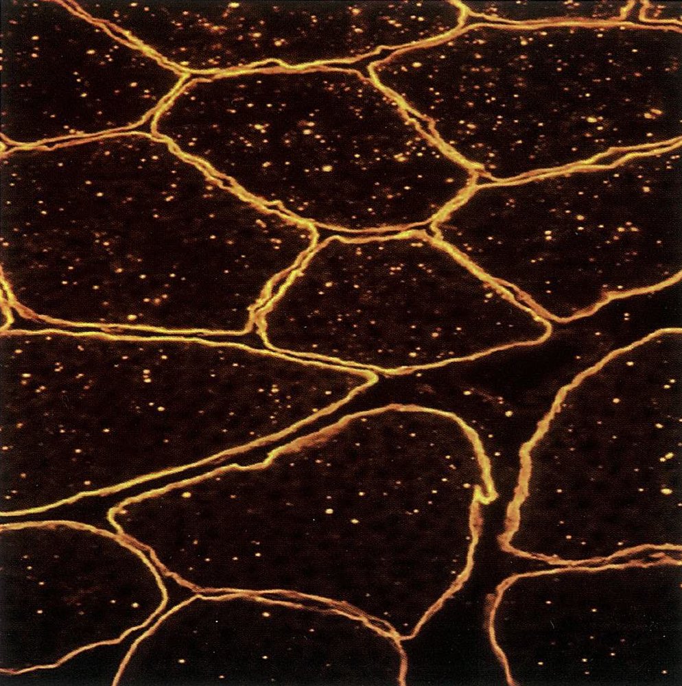
Dystrophin immunostaining shows the difference at tissue level. Normal muscle has a continuous ring of dystrophin around every fiber, while in DMD the stain is almost completely absent. BMD falls in between, with reduced, patchy staining.
| Duchenne vs. Becker muscular dystrophy | ||
|---|---|---|
| Feature | Duchenne (DMD) | Becker (BMD) |
| Genetics & chromosome | X-linked recessive; dystrophin gene at Xp21; usually a frameshift deletion | X-linked recessive; same gene at Xp21; usually an in-frame deletion |
| Dystrophin protein | Absent or severely deficient (Deleted) | Present but reduced/defective (Better) |
| Age of onset | 2–3 years (range 2–6) | 5–15 years |
| Severity & presentation | Progressive proximal weakness, Gowers sign, calf pseudohypertrophy, waddling gait, toe walking | Same pattern but milder and slower; proximal muscles |
| Ambulation | Loses walking by ~12 years; wheelchair-dependent by adolescence | Still walking after 15 years |
| Comorbidities | Dilated cardiomyopathy, scoliosis, respiratory muscle weakness (aspiration, infection) | Cardiomyopathy (the dominant problem) |
| Prognosis | Death at 20–30 years from respiratory or heart failure | Death at 40–50 years (4th–5th decade) from heart failure |
Key Points for Exams
- GBS = ascending, symmetric flaccid paralysis + areflexia 1–4 weeks after an infection. Campylobacter jejuni is the most common trigger.
- GBS is caused by molecular mimicry against Schwann cell antigens, demyelinating the ventral roots and peripheral nerves.
- In GBS the Babinski is downgoing, bladder is usually normal, pupils are spared, and the child is afebrile. A sensory level or early sphincter loss suggests a cord lesion.
- Bilateral facial weakness (CN VII) occurs in 40–50% of GBS.
- Miller Fisher = ophthalmoplegia + ataxia + areflexia (anti-GQ1b).
- CSF: ↑ protein, normal cells (albuminocytologic dissociation), appearing about 1 week after onset. EMG/NCS: slowed conduction or conduction block.
- The first priority in GBS is respiratory monitoring (FVC). Treat with IVIG or plasmapheresis; steroids do not help.
- Children with GBS usually recover completely.
- DMD: X-linked recessive, dystrophin (Xp21) absent. Boys aged 2–3 present with Gowers sign, calf pseudohypertrophy, waddling gait and toe walking.
- DMD workup: CK first → genetic testing confirms → biopsy only if genetics is negative. Raised AST/ALT come from muscle, not liver.
- At DMD diagnosis, get an echocardiogram and ECG (dilated cardiomyopathy). Glucocorticoids delay wheelchair use.
- In DMD, walking is lost by about 12 years; death at 20–30 years from respiratory or heart failure.
- BMD: reduced dystrophin, onset 5–15 years, still walking after 15, cardiomyopathy dominant, death at 40–50 years from heart failure.
احصل على التجربة الكاملة
اشترك للوصول لفيديوهات الشرح التفصيلي والبطاقات التعليمية التفاعلية وأسئلة الممارسة مع تتبع التقدم.