شرح المدرسين

Summary
Malrotation is a congenital failure of normal embryologic rotation of the midgut (occurring around week 10 of gestation) that results in abnormal bowel positioning and formation of fibrous peritoneal bands called Ladd bands. The most serious complication is midgut volvulus—twisting of the bowel around its superior mesenteric artery (SMA) blood supply—which presents as a surgical emergency, particularly in neonates presenting with bilious vomiting within the first week of life. Early recognition and imaging (upper GI series) are critical for diagnosis, as delayed treatment leads to bowel ischemia, necrosis, perforation, and sepsis within hours. While colonic volvulus (sigmoid and cecal) occurs in older adults with chronic constipation, midgut volvulus predominantly affects infants and young children, with 80% of cases occurring before age 1 year. Definitive treatment is surgical: the Ladd procedure (untwisting, division of Ladd bands, and fixation of bowel) for malrotation with volvulus.
Embryology & Pathophysiology
Normal Embryologic Development of the Midgut
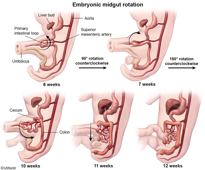
During weeks 6–12 of gestation, the primitive midgut undergoes critical counterclockwise rotation of approximately 270° around the superior mesenteric artery (SMA) as its anatomic axis:
- Week 6: Midgut herniates out of the abdomen into the umbilical cord
- Week 7: 90° counterclockwise rotation begins as bowel extends into the cord
- Week 10: Additional 180° rotation completes the 270° total arc
- Weeks 10–12: Bowel returns to the abdomen and becomes fixed by peritoneal attachments—the duodenum at the ligament of Treitz (should be left of midline at L2 level) and the ileocecal junction in the right lower quadrant
Malrotation: Failure of Normal Rotation
Malrotation occurs when the normal counterclockwise rotation process is interrupted or incomplete, resulting in abnormal anatomic variants:
- Nonrotation: Small bowel remains entirely on the right side; colon entirely on the left side
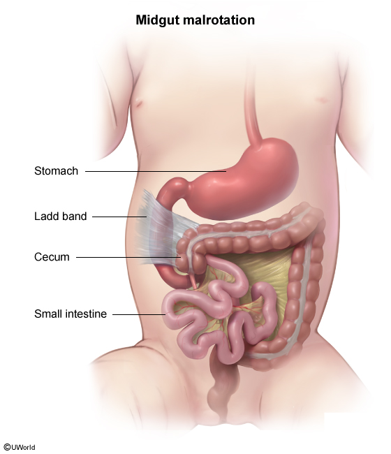
- Incomplete rotation: Cecum and ascending colon remain fixed in the right upper quadrant by fibrous peritoneal bands
Ladd Bands & Risk of Obstruction and Volvulus
Because the malrotated bowel lacks normal peritoneal fixation attachments, Ladd bands form—abnormal fibrous peritoneal tissues—that:
- Compress the duodenum (especially the third and fourth portions), causing partial or complete intestinal obstruction even without volvulus
- Create an abnormally narrow mesenteric base around the SMA, predisposing the bowel to twist
- Allow volvulus to occur: The bowel twists around the SMA, acutely occluding arterial inflow and venous outflow, leading to ischemia within 2–4 hours
Epidemiology & Associated Congenital Anomalies
Incidence & Age of Presentation
- Malrotation incidence: 1 in 500 live births; slight male predominance
- Midgut volvulus: 80% of cases occur before age 1 year (peak: <6 months); can present at any age but rare after infancy
- Colonic volvulus (sigmoid/cecal): More common in older adults (typically >60 years) with chronic constipation or neurologic disorders causing dysmotility
Critical Clinical Pearl
Malrotation is present at birth in 1 in 500 infants, but volvulus is not inevitable. Only a subset of malrotated infants develop acute volvulus; others may have asymptomatic malrotation discovered incidentally on imaging done for other reasons. However, any malrotated infant presenting with bilious vomiting must be assumed to have volvulus until proven otherwise.
Associated Congenital Anomalies
Malrotation frequently co-occurs with other congenital abnormalities, suggesting a common embryologic disturbance. The most commonly tested associations are:
- Abdominal wall defects: Gastroschisis (defect to right of umbilicus), omphalocele (midline fascial defect)
- Situs inversus totalis or heterotaxy: Complete or partial mirror-image anatomy; abnormal left-right organ laterality
- Cardiovascular defects: Transposition of the great arteries (TGA), tetralogy of Fallot, other cyanotic heart disease
- Hirschsprung disease: Aganglionic bowel segment predisposing to obstruction; may coexist with malrotation
Clinical Features
Acute Presentation: Midgut Volvulus
Neonates & Infants (typically ≤1 year):
- Bilious vomiting (the hallmark finding—vomitus is green/yellow from bile, never purely nonbilious)
- Onset typically within first 2 weeks of life; sudden-onset colicky abdominal pain
- Abdominal distention and visible peristaltic waves
- Blood-tinged or "currant jelly" stools (late, concerning sign indicating bowel ischemia)
- Irritability, lethargy, dehydration, and signs of shock if bowel is necrotic
Older Children & Adults (if malrotation undiagnosed until later in life):
- Chronic intermittent symptoms: recurrent nonbilious vomiting, crampy abdominal pain, change in bowel habits, failure to thrive
- Acute exacerbation: sudden severe pain, bilious emesis, obstruction
- Abdominal distention, tympany on percussion
Physical Examination Findings
- Early stage: Often deceptively normal or mild tenderness only—do NOT be falsely reassured by a benign initial exam
- Progressive findings: Abdominal distention, visible peristalsis, absence of bowel sounds (ileus)
- Late/severe stage: Peritonitis (rigid abdomen, guarding, rebound tenderness), signs of septic shock (fever, tachycardia, hypotension, altered mental status)
Differential Diagnosis in the Neonatal/Infantile Period
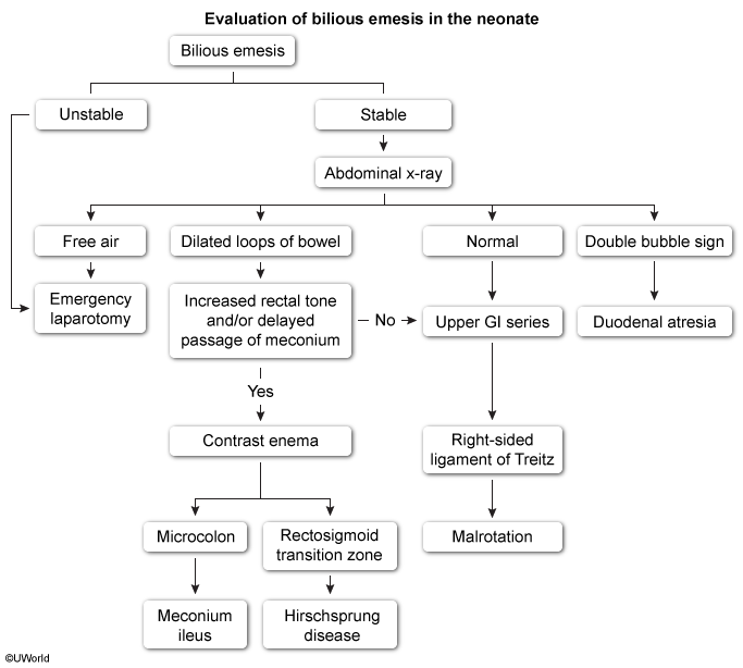
While other causes of bilious vomiting exist, malrotation with volvulus must be the leading hypothesis in any neonate or young infant with bile-stained vomitus:
- Duodenal atresia: Presents with the classic "double bubble" sign on X-ray (air in stomach and duodenum only) and feeding intolerance starting immediately after birth; usually nonbilious or mildly bilious
- Intussusception: Peak age 6–18 months; presents with episodic colicky pain and "currant jelly" stools; often has palpable "sausage" mass in the right upper quadrant
- Intestinal atresia (jejunal/ileal): Apple-peel type (Type IIIb) from in utero vascular occlusion; similar presentation with bilious vomiting
- Necrotizing enterocolitis (NEC): Preterm infants; pneumatosis intestinalis on X-ray
Diagnosis & Imaging
Initial Plain Radiography (Abdominal X-ray)
Sensitivity: Low (often nonspecific); may be entirely normal in early malrotation without volvulus
- Nonspecific findings: Multiple air-fluid levels (suggesting bowel obstruction), dilated small bowel loops, loss of haustra (haustra are the normal indentations in colon), abdominal distention
- Concerning/emergent findings:
- Pneumoperitoneum (free air under the diaphragm on upright or left lateral decubitus film) → bowel perforation, absolute surgical emergency
- Ground-glass appearance → ascites (suggests bowel ischemia with peritoneal fluid)
Gold Standard: Upper Gastrointestinal Series with Barium or Water-Soluble Contrast
This is the definitive diagnostic test for malrotation. Timing is critical: if volvulus is suspected clinically and the patient is unstable, proceed directly to surgical consultation and OR without waiting for upper GI series.
Key diagnostic findings for malrotation (even without active volvulus):
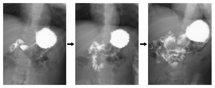
- Failure of the duodenum to cross the midline → duodenum positioned abnormally to the right of the vertebral column; normally the duodenum crosses to the left at the level of the duodenojejunal (DJ) flexion
- Abnormal position of the duodenojejunal flexion (Ligament of Treitz) → should be at the left of midline at approximately the L2 vertebral level; if right-sided, too low, or absent, malrotation is confirmed
- Small bowel loops abnormally positioned to the right side of the abdomen (normally they occupy the central and left abdomen)
Pathognomonic Finding: The "Corkscrew" Sign (Active Volvulus)
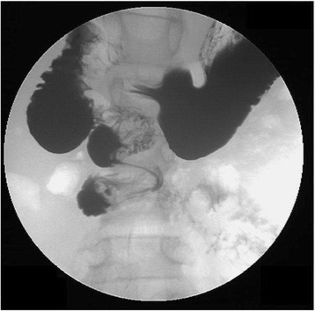
When the malrotated bowel twists around the SMA (volvulus), a characteristic appearance is seen:
- Corkscrew or spiral sign: Twisted appearance of the distal duodenum and proximal jejunum, spiraling as the bowel rotates around the SMA axis
- This sign is pathognomonic for midgut volvulus and mandates immediate surgical exploration
Additional Imaging Modalities
- CT abdomen/pelvis: May show the mesenteric "whirl" sign (rotation of mesentery and vessels around the SMA), though CT is less commonly the first test if clinical suspicion is high. Useful in equivocal cases or when complications (perforation, necrosis extent) need assessment
- Ultrasound: In experienced hands, can detect reversed relationship of SMA (should be left of SMV) and right-sided small bowel; useful in some centers as a first-line modality, especially in neonates to avoid radiation
- Barium enema: May show the cecum in an abnormal (high, right-sided) position; less commonly used in modern practice but can be adjunctive
Management & The Ladd Procedure
The following table presents a unified, side-by-side comparison of management approaches across the three volvulus types:
| Comparative Overview: Midgut, Sigmoid, and Cecal Volvulus | |||
|---|---|---|---|
| Parameter | Midgut Volvulus | Sigmoid Volvulus | Cecal Volvulus |
| Age & Epidemiology | Neonates & infants <6 months (80% before age 1); both sexes | Elderly (>60 years); chronic constipation, Parkinson's, neuroleptics | Middle-aged to elderly; female predominance; history of chronic constipation |
| Presentation | Acute: bilious vomiting, abdominal distention, colicky pain; onset within first 2 weeks of life | Acute or chronic intermittent; severe colicky pain, constipation, abdominal distention | Acute or intermittent; right-sided pain, abdominal distention, constipation |
| Etiology | Malrotation with narrow mesenteric base predisposing to twisting around SMA | Redundant sigmoid colon & elongated sigmoid mesentery; twist around its own mesentery | Ileocecal ligament laxity; cecal prolapse into pelvis; twist around ileocolic vessels |
| Imaging Gold Standard | Upper GI series: corkscrew sign (spiral distal duodenum/proximal jejunum); failure of duodenum to cross midline | Abdominal X-ray: coffee-bean sign (apex of dilated sigmoid points toward left flank); 'bird's beak' on barium enema | CT or abdominal X-ray: transition point at hepatic flexure; dilated right colon; dilated terminal ileum |
| Initial Management | Emergent surgical exploration + Ladd procedure (untwist, divide bands, broaden mesentery, reposition, appendicectomy); no non-operative option | Try endoscopic detorsion (flexible sigmoidoscopy) first; >90% success; avoid perforation risk; if fails → colectomy | Surgical reduction or right hemicolectomy; higher recurrence if not resected |
| Surgical Urgency | EMERGENCY: ischemia within 2–4 hours; do NOT delay for confirmatory imaging if unstable | Urgent but permits trial of endoscopy if stable & no perforation signs | Urgent; lower recurrence if resected; elective colectomy if no perforation |
| Mortality if Delayed >24 hours | Increases from <5% to 10–30%; extent of necrosis determines morbidity | 10–20% if perforation; usually lower if managed early | Lower than midgut (less time-critical); depends on extent of necrosis |
Pre-operative Stabilization (Before Laparotomy)
- Nothing by mouth (NPO) to prevent aspiration
- Nasogastric (NG) tube placement (decompresses stomach and proximal bowel, reducing risk of aspiration during induction)
- Two large-bore IVs + aggressive fluid resuscitation: Bowel obstruction causes massive fluid sequestration into the bowel lumen and peritoneal cavity; typical deficit is 50–100 mL/kg of isotonic crystalloid over first 1–2 hours, then maintenance
- Broad-spectrum IV antibiotics (to cover gram-positive, gram-negative, and anaerobic bacteria given risk of perforation and bacterial translocation):
- Ampicillin 50 mg/kg/dose IV Q6H + Gentamicin 7.5 mg/kg/dose IV Q8H (or once-daily dosing) + Clindamycin 15 mg/kg/dose IV Q6H
- Alternative: 3rd-generation cephalosporin (ceftriaxone or cefotaxime) + clindamycin
- Correction of electrolyte abnormalities: Hypokalemia, hyponatremia, hypochloremia from vomiting
- Blood cultures if febrile (risk of sepsis)
- Immediate surgical consultation (do NOT delay for imaging if unstable)
Operative Steps of the Ladd Procedure
- Open abdomen via midline laparotomy
- Untwist the volvulus: Gentle counterclockwise (opposite direction of the twist) rotation of the entire midgut mass to unwinding the volvulus around the SMA
- Assess bowel viability:
- Inspect for signs of ischemia (dark purple/black color, lack of bleeding when incised, no peristalsis)—viable bowel is pink/red and bleeds
- If necrosis is limited (small segment), proceed with resection; if extensive, consider staged approach or limitation of resection in critically ill infants
- Divide Ladd bands: Carefully divide the fibrous peritoneal bands overlying the duodenum (third and fourth portions) to relieve duodenal obstruction; extend division laterally
- Broaden the mesenteric base: Divide peritoneum lateral to the duodenum and along the right colon mesentery, widening the angle of the SMA mesenteric pedicle to prevent recurrent volvulus; the goal is a >90° angle between the SMA origin and the mesentery
- Reposition the bowel: Place small bowel loops on the right side of abdomen and colon on the left side (reversed from normal but appropriate positioning after malrotation correction)
- Fixation (peritoneal fixation, not suturing): Some surgeons fix the duodenum laterally to the right peritoneum and the ileocecal junction to prevent recurrence; pure adhesiolysis without fixation is acceptable if dissection was adequate
- Appendicectomy: Prophylactic appendicectomy may be performed to prevent future confusion in diagnosis of appendicitis (cecum is in an unusual location post-Ladd)
- Close abdomen: Perform careful hemostasis and close in layers
Post-operative Management
- Continue broad-spectrum antibiotics for 7–10 days (or longer if perforation occurred)
- NG tube to low intermittent suction for 1–3 days until bowel function returns (flatus/stool passage)
- Gradual diet advancement: Start with clear liquids, advance as tolerated once bowel sounds return and emesis resolves
- Monitor for complications: Anastomotic leak (rare unless resection was performed), recurrent obstruction from adhesions, sepsis
Outcomes After Ladd Procedure
- Recurrence of volvulus: Rare (1–3%) if Ladd bands are completely divided and mesentery is sufficiently broadened
- Short bowel syndrome: Only if extensive necrosis required resection of >50% of small bowel; rare with prompt surgery
- Secondary adhesions: Uncommon cause of obstruction after Ladd procedure when surgery was uncomplicated
- Overall survival: >95% if surgery is performed before irreversible bowel necrosis; mortality increases significantly if diagnosis is delayed >24 hours or massive bowel resection was needed
Operative Mnemonic for the Ladd Procedure
| Ladd Procedure Operative Mnemonic – جملة تذكرية | |
UDBRA = Remember the operative sequence for fixing malrotation with volvulus:
Hospital pearl: تذكر UDBRA كأنك بتـ undo/redo الـ malrotation من الأول. |
جملة تذكرية |
Key Points for Exams – نقاط مهمة للامتحانات
High-Yield Epidemiology & Embryology
- Malrotation incidence: 1 in 500 live births; male predominance
- Peak presentation of midgut volvulus: Neonates and infants <6 months old; 80% of cases <1 year
- Normal midgut rotation: Occurs week 10 of gestation; 270° counterclockwise around SMA axis
- Malrotation at birth: Present in 1 in 500 infants, but only a fraction develop symptomatic volvulus
Hallmark Clinical Feature
- Bilious vomiting in a neonate = midgut volvulus until proven otherwise (highest-yield fact for exams)
- NOT feeding intolerance, NOT reflux, NOT gastroenteritis—bilious vomitus indicates bile has refluxed from below the pylorus, implying distal obstruction
Imaging Gold Standard & Diagnostic Criteria
- Upper GI series with barium: Gold standard diagnostic test for malrotation
- Key finding for malrotation: Duodenum fails to cross midline; duodenojejunal flexion (Ligament of Treitz) positioned right of midline instead of left
- Pathognomonic finding for volvulus: Corkscrew (spiral) sign in distal duodenum/proximal jejunum
- Distinction between volvulus types: Corkscrew (small bowel, neonates) vs. coffee bean (sigmoid colon, elderly)
Management Essentials
- Midgut volvulus = surgical emergency: Do NOT attempt non-operative management; do NOT observe with serial exams
- Pre-operative stabilization: NPO, NG tube, aggressive IV fluids (50–100 mL/kg bolus), broad-spectrum antibiotics (amp/gent/clinda), electrolyte correction
- Definitive procedure: Ladd procedure (UDBRA: Untwist, Divide bands, Broaden mesentery, Reposition, Appendicectomy)
- Sigmoid volvulus (adult): Try endoscopic detorsion (flexible sigmoidoscopy) first; elective colectomy if recurrence or perforation
- Cecal volvulus: Surgical reduction or right colectomy
Acute Complications of Untreated Volvulus
- Bowel ischemia and necrosis: Twisting of the mesentery occludes the SMA (and superior mesenteric vein, SMV) perfusion to the midgut. Without restoration of blood flow within 2–4 hours, transmural (full-thickness) infarction develops. Ischemic bowel becomes dark purple/black, loses contractility, and no longer maintains the intestinal barrier.
- Perforation: Necrotic bowel wall perforates, spilling intestinal contents (bacteria, enzymes, stool) into the peritoneal cavity.
- Peritonitis and sepsis: Bacterial translocation through the damaged bowel and peritoneal inflammation trigger a systemic inflammatory response. If untreated, septic shock and multi-organ failure (renal failure, coagulopathy, respiratory failure) develop rapidly.
- Hypovolemic shock: Massive fluid sequestration into the obstructed bowel lumen, third-space fluid shifts into the peritoneal cavity, and ongoing vomiting cause severe dehydration. Shock can be present before perforation occurs, especially in delayed presentations.
Late Complications
- Short bowel syndrome: If volvulus was diagnosed late and extensive bowel necrosis required resection of >50–60% of the small bowel, the remaining bowel may have insufficient absorptive surface area, leading to chronic diarrhea, malabsorption, and failure to thrive. Long-term parenteral nutrition or small bowel transplantation may be needed.
- Adhesions: Post-surgical adhesions can cause secondary intestinal obstruction weeks to months after the Ladd procedure, though this is uncommon if the initial surgery was uncomplicated.
Prognosis
- With early diagnosis and treatment (surgery <12–24 hours): Survival >95%; most infants have excellent long-term outcomes with normal bowel function
- Delayed diagnosis (>24–48 hours): Mortality increases to 10–30% depending on extent of necrosis; survivors often have significant morbidity (short bowel, failure to thrive)
- Post-operative recurrence: Rare (1–3%) after proper Ladd procedure; if recurrence occurs, reoperation is needed
Classic USMLE/JMC Board-Exam Vignette
"A 5-day-old full-term male neonate presents to the ED with green bilious vomiting and abdominal distention. On exam, the infant is irritable but the abdomen is soft without obvious mass. Plain abdominal X-ray shows dilated loops of small bowel with air-fluid levels but no pneumoperitoneum. What is the next best step in management?"
Answer: Emergent upper GI series with water-soluble contrast (or, if the infant is hemodynamically unstable, proceed directly to surgical consultation without waiting for imaging). The goal is to identify the corkscrew sign and confirm malrotation with volvulus before ischemia progresses. If volvulus is confirmed, emergency laparotomy and Ladd procedure.
Do NOT Miss
- Bilious vomiting is never normal in a neonate—always investigate
- A soft, benign-feeling abdomen does NOT rule out volvulus—the infant can look deceptively well early in the disease
- Delay in diagnosis of >24 hours increases mortality from <5% to >10–20%
- Pneumoperitoneum (free air on X-ray) = perforation = absolute surgical emergency
Common Exam Pitfalls
- Confusing nonbilious vomiting (pyloric stenosis, reflux) with bilious vomiting (obstruction below pylorus)
- Assuming normal or soft abdomen = not surgical—ischemic bowel can be silent early
- Delaying surgical consultation for "one more X-ray" or "observation"—volvulus is time-sensitive
- Confusing corkscrew (midgut, emergency) with coffee bean (sigmoid, can try endoscopy)—age and location of twist are key
احصل على التجربة الكاملة
اشترك للوصول لفيديوهات الشرح التفصيلي والبطاقات التعليمية التفاعلية وأسئلة الممارسة مع تتبع التقدم.