شرح المدرسين

SUMMARY
Hirschsprung's disease (congenital aganglionic megacolon) is a developmental disorder defined by the absence of ganglion cells in the Auerbach (myenteric) and Meissner (submucosal) plexuses of the distal colon, arising from failed craniocaudal migration of neural crest cells.
The aganglionic segment remains tonically contracted, producing a functional obstruction with proximal colonic dilation. Neonates classically present with bilious vomiting, abdominal distension, and failure to pass meconium within the first 48 hours; older children present with chronic constipation and failure to thrive. Diagnosis is confirmed by rectal suction biopsy (gold standard) showing absent ganglion cells, and definitive treatment is surgical resection of the aganglionic segment.
Pathophysiology & Etiology
Hirschsprung's disease is a congenital aganglionic megacolon defined by the absence of enteric ganglion cells in both the Auerbach (myenteric) and Meissner (submucosal) plexuses of the distal colon. It results from failed craniocaudal (caudal) migration of neural crest cells along the developing gut, leaving the most distal bowel without intramural ganglia.
Because the aganglionic segment cannot relax, it remains in a state of persistent tonic contraction, creating a functional obstruction. Stool accumulates behind this narrowed segment, producing progressive proximal dilation (megacolon). The rectosigmoid is the most commonly affected level.
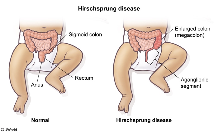
Epidemiology: the disease is roughly five times more common in male infants, and up to 80% of cases have a positive family history.
Associations (high-yield): the disorder is classically linked with Down syndrome, Chagas' disease (in which amastigotes destroy ganglion cells), and loss-of-function mutations in RET. See the Down syndrome comorbidities by body system for the other GI, cardiac, and haematologic anomalies that cluster with trisomy 21.
| Mnemonic – جملة تذكرية | |
“Dr. Chagas” has no ganglion cells — the three classic associations of Hirschsprung disease:
|
جملة تذكرية |
Clinical Presentation
Presentation depends on the patient's age and on the length of the aganglionic segment.
Neonates typically show:
- Failure to pass meconium within the first 48 hours of life
- Bilious vomiting
- Abdominal distension with signs of large bowel obstruction
- ± Enterocolitis (fever, explosive diarrhoea, sepsis)
| Mnemonic – جملة تذكرية | |
|
The neonate is a “BAD baby” — the classic triad of Hirschsprung disease:
|
جملة تذكرية |
Children / adolescents (usually short-segment disease) present later with chronic constipation and failure to thrive.
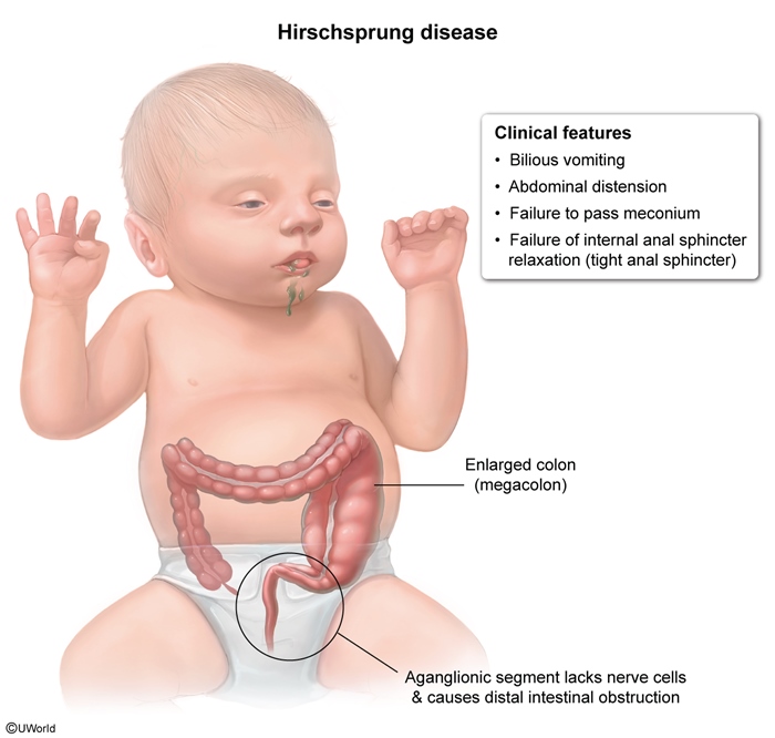
Physical examination: a distended abdomen, an extremely tight anal sphincter, and an empty rectal vault (no stool on digital exam). Withdrawal of the examining finger triggers forceful expulsion of stool and gas — the classic “squirt sign.”
| Exam Trap - فخ امتحاني | |
|
If you find an empty rectum (empty rectal vault) on digital rectal exam, and after withdrawal of your finger stool or gas is expelled forcefully (positive squirt sign) — this is the classic picture of Hirschsprung, NOT functional constipation, because in functional constipation the rectum would be filled with stool. |
تذكر |
Diagnosis
Work-up proceeds from supportive imaging toward the confirmatory biopsy:
- Biopsy (gold standard): rectal suction biopsy demonstrating absence or paucity of ganglion cells in the submucosa confirms the diagnosis.
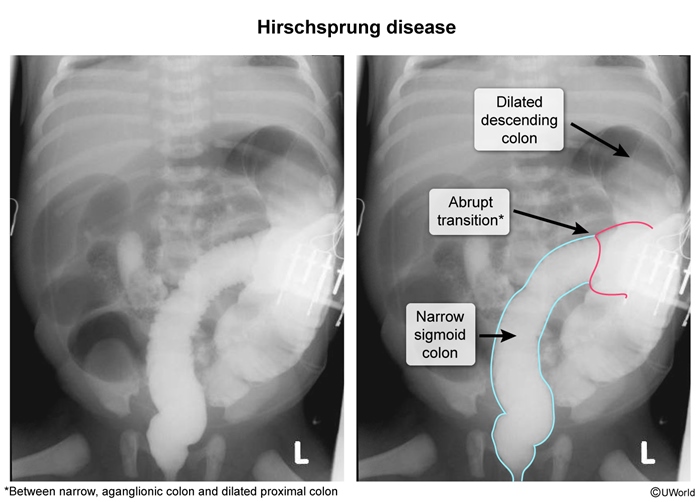
- Imaging — contrast enema (adjunct): reveals the rectosigmoid transition zone — a narrowed distal aganglionic segment with abrupt proximal dilation.
- Imaging — abdominal radiograph: distended bowel loops with absence of air in the rectum, consistent with large bowel obstruction.
- Special test — anorectal manometry (adjunct): failure of the internal anal sphincter to relax.
The transition zone is the hallmark radiographic clue on the contrast study.
| ملاحظة سريرية – Clinical Note | |
الـ rectal suction biopsy لازم تاخد عيّنة من الـ submucosa تحديداً مشان تثبت غياب الـ ganglion cells (Meissner plexus) — هاي هي الـ gold standard، والخزعة السطحية اللي ما توصل للـ submucosa ممكن تعطي نتيجة مضللة. |
ملاحظة |
Differential Diagnosis
Delayed meconium passage and neonatal large bowel obstruction have a broad differential, including:
- Imperforate anus
- Functional constipation
- Intussusception
- Duodenal atresia
- Meconium ileus
- Necrotising enterocolitis
The most important distinction is between Hirschsprung's disease and meconium ileus, separated by their associated disorder, level of obstruction, meconium consistency, and the squirt sign.
Template: Medical Comparison
| Hirschsprung Disease vs Meconium Ileus | ||
|---|---|---|
| Feature | Hirschsprung's disease | Meconium ileus |
| Associated disorder | Down syndrome | Cystic fibrosis |
| Typical level of obstruction | Rectosigmoid (large bowel) | Ileum (small bowel) |
| Meconium consistency | Normal | Inspissated (thick, sticky) |
| “Squirt sign” | Positive | Negative |
Management & Complications
Complications:
- Enterocolitis — an increased risk in Hirschsprung's disease and the most serious, potentially life-threatening complication.
- Colonic perforation / rupture.
Management: definitive treatment is surgical resection of the aganglionic segment of colon, restoring continuity by bringing normally innervated (ganglionic) bowel down to the anus.
Key Points for Exams
نقاط مهمة للامتحانات — the highest-yield facts:
- Hirschsprung = congenital aganglionosis (absent Auerbach + Meissner plexuses) from failed neural crest cell migration.
- Classic neonate: bilious vomiting, abdominal distension, failure to pass meconium >48 h.
- Physical exam: tight anal sphincter, empty rectal vault, positive squirt sign.
- Rectosigmoid = most common level; rectal suction biopsy (submucosa) = gold standard.
- Associations: Down syndrome, RET (loss-of-function), Chagas disease.
- Male predominance (~5:1); ~80% positive family history.
- Most feared complication: enterocolitis; definitive treatment is surgical resection of the aganglionic segment.
| Important – فكرة سؤال | |
Gold standard vs adjunct: the rectal suction biopsy (absent ganglion cells in the submucosa) confirms the diagnosis. A contrast enema showing the rectosigmoid transition zone and anorectal manometry are only supportive adjuncts — never choose imaging over biopsy for definitive diagnosis. |
تذكر |
| Mnemonic – جملة تذكرية | |
“BAD baby” = Bilious vomiting, Abdominal distension, Delayed meconium (>48 h). “Dr. Chagas” = Down syndrome, RET mutation, Chagas disease — the three key associations. |
جملة تذكرية |
احصل على التجربة الكاملة
اشترك للوصول لفيديوهات الشرح التفصيلي والبطاقات التعليمية التفاعلية وأسئلة الممارسة مع تتبع التقدم.