Summary
Disorders of B lymphocytes are the most common primary immunodeficiency. Maternal IgG protects infants; symptoms typically emerge after 6 months. Recurrent sinopulmonary infection with encapsulated bacteria, chronic Giardia diarrhea, and dangerous enteroviral infection (in XLA) are characteristic. Four main types differ by B-cell count and immunoglobulin pattern: XLA (no B cells, all immunoglobulins low); CVID (normal B cells, low IgG/IgA/IgM, adult onset); selective IgA deficiency (isolated low IgA, usually asymptomatic); hyper-IgM syndrome (high IgM, low IgG/IgA/IgE, opportunistic infections). Workup includes immunoglobulin quantitation, vaccine titers, and flow cytometry. Treatment: lifelong immunoglobulin replacement or stem cell transplantation. Live vaccines are contraindicated; IgA-containing blood products can cause anaphylaxis in IgA deficiency.
Overview and Laboratory Comparison
B-cell (humoral) defects are the most common category of primary immunodeficiency. Antibody production fails, so the patient loses opsonization, toxin and virus neutralization, and mucosal IgA protection, while T-cell immunity is usually preserved. The warning signs and general classification of immunodeficiencies are covered in the Immunology overview lesson.
Shared features
- Age of onset: usually after 4–6 months, once transplacental maternal IgG has been used up; CVID typically appears after puberty.
- Typical pathogens:
- Encapsulated bacteria (S. pneumoniae, H. influenzae type b) → recurrent otitis media, sinusitis and pneumonia; also bacteremia and meningitis.
- Giardia lamblia → chronic diarrhea and malabsorption (loss of secretory IgA).
- Enteroviruses → chronic meningoencephalitis, characteristic of XLA.
- Hallmark labs: low immunoglobulins and a poor antibody response to vaccines.
- Course: infections respond to antibiotics but keep coming back. Severe viral or fungal infections suggest a T-cell or combined defect instead; hyper-IgM syndrome is the exception in this group.
Diagnostic approach
- Step 1: Quantitative serum IgG, IgA and IgM (± IgE) is the best initial test for recurrent sinopulmonary infection. Interpret the results against age-specific ranges.
- Step 2: Vaccine titers show whether the antibody defect is functional.
- Step 3: Flow cytometry for CD19+ B cells and CD3+ T cells. B cells are absent in XLA but normal in CVID and hyper-IgM. When IgM is high, test CD40 ligand expression on activated CD4+ T cells.
- Step 4: Genetic testing (eg, BTK, CD40LG) is confirmatory and guides family counseling.
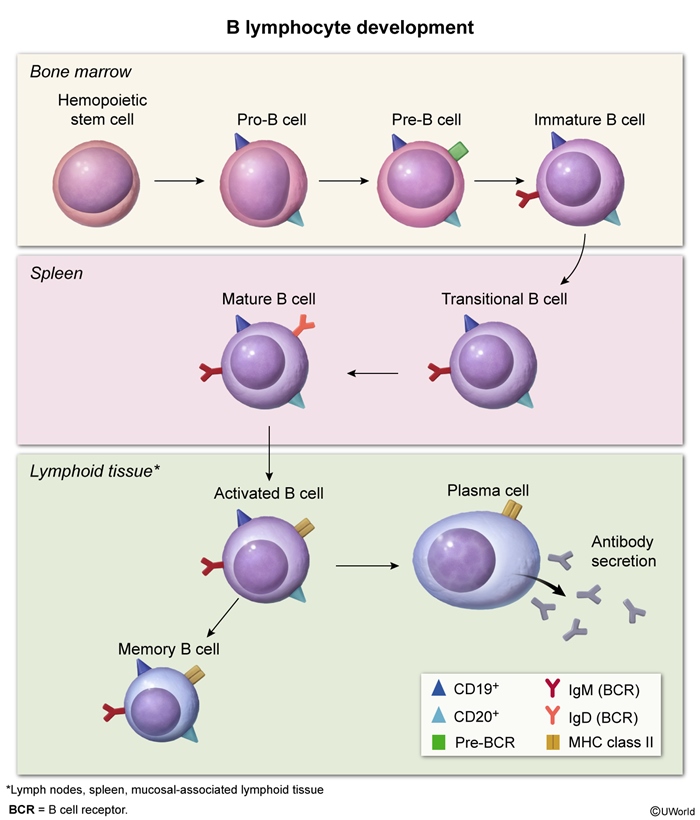
Each disorder has its own pattern of B-cell count and immunoglobulin classes:
| Primary humoral immunodeficiencies: B-cell count and immunoglobulin pattern | ||||||
|---|---|---|---|---|---|---|
| Disorder | B-cell count (CD19+) | IgG | IgA | IgM | IgE | Distinguishing clue |
| X-linked agammaglobulinemia | ↓↓ / absent | ↓ | ↓ | ↓ | ↓ | Boy >6 months, no tonsils, enteroviral meningoencephalitis |
| Common variable immunodeficiency | Normal | ↓↓ | ↓ | ↓ | ↓ | Onset after puberty (20–40 yr); autoimmunity, granulomas, lymphoma |
| Selective IgA deficiency | Normal | Normal | ↓↓ (<7 mg/dL) | Normal | Normal | Usually asymptomatic; celiac/atopy; transfusion anaphylaxis |
| Hyper-IgM syndrome (CD40L deficiency) | Normal | ↓ | ↓ | Normal or ↑ | ↓ | Infant boy with Pneumocystis pneumonia, Cryptosporidium, neutropenia |
| Hyper-IgE syndrome (for contrast) | Normal | Normal | Normal | Normal | ↑↑ |
Cold staphylococcal abscesses, eczema; a STAT3/Th17–neutrophil defect, not a B-cell defect |
X-Linked Agammaglobulinemia (XLA)
Pathophysiology
- Genetics: an X-linked recessive mutation in the BTK gene (Xq22). This gene encodes Bruton tyrosine kinase, which carries signals from the pre-B-cell receptor.
- Maturation arrest: B-cell precursors in the bone marrow are normal, but development stops at the pre-B stage.
- Consequences: almost no circulating mature (CD19+) B cells, no plasma cells, no lymphoid follicles or germinal centers, and very low levels of every immunoglobulin class. T cells are normal in number and function.
Clinical features
- Who and when: boys who present in the second half of the first year. Some are recognized only after age 1, once they have needed many courses of antibiotics.
| فخ امتحاني | |
| المريض بيجي بعد 6 شهور مش قبل، لأن الـ maternal IgG بتكون لسا حاميته بالبداية. | تذكر |
- Recurrent pyogenic infections: otitis media, sinusitis and pneumonia from S. pneumoniae and H. influenzae; also sepsis, meningitis and osteomyelitis. Invasive Hib disease (eg, epiglottitis) in a fully vaccinated boy is a classic clue.
- Chronic enteroviral infection (meningoencephalitis) is the most feared viral complication.
- Persistent giardiasis, with diarrhea and poor growth.
- Examination: small or absent tonsils, adenoids and lymph nodes. This is the only characteristic physical finding.
- Not a feature: severe EBV mononucleosis, fungal or opportunistic infections, because T cells are intact.
Diagnosis
- Quantitative immunoglobulins: IgG, IgA, IgM and IgE are all markedly low, and vaccine titers are absent.
- Flow cytometry (key test): CD19+ B cells are absent or nearly absent, and CD3+ T cells are normal.
- CBC: may look normal, because B cells make up only a small share of blood lymphocytes.
- Most accurate (confirmatory) test: genetic testing showing a BTK mutation.
Management
- First-line, lifelong: immunoglobulin replacement. Give IVIG 400–600 mg/kg every 3–4 weeks or subcutaneous IgG 100–200 mg/kg weekly, and adjust the dose to the clinical response.
- Infections: start broad-spectrum antibiotics early. Add prophylactic antibiotics if infections continue despite adequate IgG.
- Vaccines: live vaccines are contraindicated (oral polio, MMR/MMRV, BCG, yellow fever, live intranasal influenza, oral typhoid). Inactivated vaccines are safe but produce little antibody. IVIG also neutralizes the measles vaccine virus.
| Exam trap | |
| Do not give live polio vaccine to an XLA patient because he is at high risk for severe enterovirus infection. | تذكر |
- Prognosis: near-normal life with regular IgG. The long-term risks are bronchiectasis and chronic enteroviral CNS disease.
The XLA summary table condenses these points for quick review.
| Mnemonic – جملة تذكرية: the 6 B's of Bruton | |
|
جملة تذكرية |
Common Variable Immunodeficiency (CVID)
Pathophysiology
- Defect: B cells are present in normal numbers but fail to become class-switched, long-lived plasma cells, and memory B cells are reduced. The result is low antibody levels with poor function.
- Genetics: about 90% of cases are sporadic, and a causative gene is found in only a minority. Males and females are equally affected; some cases run in families.
Clinical features
- Onset: classically at age 20–40, but it can start at puberty; it is rare in young children.
- Infections: recurrent sinopulmonary infections with encapsulated bacteria, and GI infections (Giardia, Salmonella, Campylobacter).
- Chronic lung disease (about one third of patients): bronchiectasis from repeated infection, or interstitial and granulomatous lung disease.
- Autoimmunity (about one quarter): most often immune thrombocytopenia and autoimmune hemolytic anemia; also pernicious anemia, rheumatoid arthritis/polyarthritis and thyroiditis.
- Granulomatous disease: sarcoid-like noncaseating granulomas in the lung, liver, spleen, lymph nodes and skin.
- GI disease: sprue-like malabsorption, IBD-like colitis, and chronic diarrhea with weight loss.
- Malignancy: lymphoma and gastric cancer (about 10% combined).
Diagnosis
- Best initial test: quantitative immunoglobulins. IgG is markedly low (≥2 SD below the mean for age; often <400 mg/dL), together with low IgA and/or IgM.
- Required: a poor response to both protein vaccines (tetanus, diphtheria) and polysaccharide vaccines (pneumococcal).
- Flow cytometry: normal total B and T cells with fewer switched memory B cells. It supports the diagnosis but is not required.
- Diagnosis of exclusion: first rule out secondary hypogammaglobulinemia (protein loss through the kidney or gut, glucocorticoids, antiepileptics, lymphoid malignancy). In children under about 4–6 years, also rule out transient hypogammaglobulinemia of infancy.
| Important – فكرة سؤال: CVID or XLA? | |
| Both have low IgG, IgA and IgM with absent vaccine responses. Flow cytometry decides: CD19+ B cells are absent in XLA but normal in CVID. Age and sex help too — XLA is an infant boy after 6 months; CVID is an adolescent or adult of either sex whose childhood was largely infection-free. | تذكر |
Management
- First-line, lifelong: immunoglobulin replacement, dosed as in XLA. It also slows the progression of chronic lung disease.
- Antibiotics: start early for each infection; use prophylaxis if infections recur despite IgG.
- Vaccines: live vaccines are contraindicated; inactivated vaccines are still given.
- Surveillance: watch for autoimmune and granulomatous disease, lymphoma and gastric cancer. Chronic lung disease and malignancy are now the leading causes of death.
The CVID summary table gives a one-glance review.
Selective IgA Deficiency
Epidemiology and pathophysiology
- Most common primary immunodeficiency: about 1 in 100 to 1 in 1,000 people, depending on the population.
- Defect: B cells fail to mature into IgA-secreting plasma cells, so serum IgA and secretory IgA are very low. IgG, IgM, B-cell numbers and T-cell immunity are normal.
- Why most patients stay well: secretory IgM partly takes over mucosal defense.
Clinical features
- Usually asymptomatic, and often found by chance (eg, during celiac screening).
- Recurrent infections: sinopulmonary (S. pneumoniae, H. influenzae) and GI (Giardia, causing chronic diarrhea and malabsorption).
- Atopy: asthma, eczema and food allergy.
- Autoimmunity: celiac disease, SLE, thyroid disease, type 1 diabetes, juvenile idiopathic arthritis, IBD and autoimmune cytopenias.
- Anaphylactic transfusion reaction: some patients with severe deficiency make anti-IgA antibodies. Even trace IgA in donor products can then cause hypotension, angioedema and bronchospasm within seconds to minutes. The immunologic transfusion reactions table contrasts it with the other reactions.
- Course: a few patients progress to CVID over time.
Diagnosis
- Criteria: serum IgA <7 mg/dL (or undetectable) in a patient older than 4 years, with normal IgG and IgM and normal vaccine responses.
- Exclude secondary causes (eg, antiepileptic drugs, other immunodeficiencies). In younger children IgA may still rise, so repeat the test later.
- Celiac pitfall: IgA-based serology (tTG-IgA, endomysial IgA) is falsely negative. Measure total IgA and use IgG-based tests (DGP-IgG, tTG-IgG).
Management
- Supportive care: prompt antibiotics for infections, prophylactic antibiotics for frequent recurrences, and routine vaccination.
- IVIG: not needed (IgG is normal) and a relative contraindication. If it is ever required, choose a low-IgA product and premedicate with an antihistamine and/or a glucocorticoid.
- Transfusion: the patient should wear a medical alert bracelet. Give washed red cells or components from IgA-deficient donors, and never unmodified whole blood.
| ملاحظة سريرية | |
| Beware: whole blood in IgA deficiency can precipitate severe anaphylaxis—you must use washed RBCs instead. | ملاحظة |
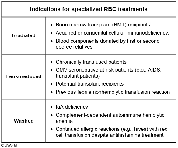
| Important – فكرة سؤال: what is NOT true in IgA deficiency | |
|
تذكر |
The IgA deficiency summary table lists the essentials in one place.
Hyper-IgM Syndrome
Pathophysiology
- Genetics: most cases are X-linked recessive mutations in CD40LG, so activated CD4+ T cells lack CD40 ligand. Rarer autosomal recessive forms involve CD40 itself or B-cell enzymes such as AID.
- B-cell arm: germinal centers and class switching need CD40L to bind CD40. Without it, B cells (normal in number) keep making IgM, and IgG, IgA and IgE stay low.
- Macrophage arm: CD40 on macrophages and dendritic cells also misses the T-cell signal. Cell-mediated immunity is weakened, which leads to opportunistic infections.
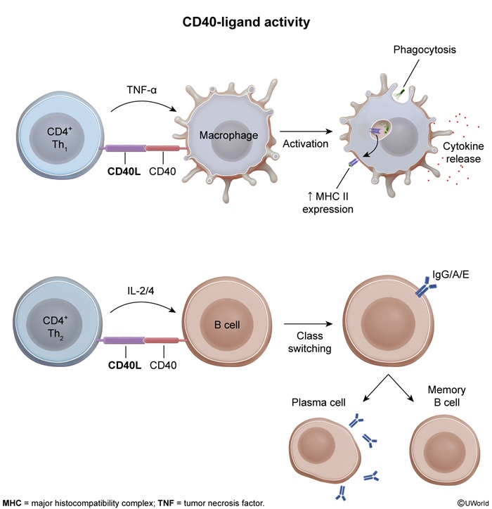
Clinical features
- Who: an infant or young boy (X-linked form), often with failure to thrive.
- Sinopulmonary infections with encapsulated bacteria, because there is no opsonizing IgG.
- Opportunistic infections: Pneumocystis jirovecii pneumonia (often the presenting infection); Cryptosporidium enteritis leading to sclerosing cholangitis and cirrhosis; CMV.
- Other infections: Giardia (no mucosal IgA), and chronic parvovirus B19 causing red-cell aplasia.
- Hematologic and autoimmune: neutropenia (common in the X-linked form) and autoimmune cytopenias.
- Examination: lymphoid tissue is usually small (no germinal centers); hepatosplenomegaly can occur with liver disease.
- Long term: a high risk of malignancy (lymphoma, liver and biliary tumors) by the second decade. The X-linked form has the worst prognosis.
Diagnosis
- Immunoglobulins: IgM normal or high; IgG, IgA and IgE low; poor vaccine titers.
- Flow cytometry: normal B- and T-cell counts with absent CD40L on activated CD4+ T cells.
- CBC: may show neutropenia, anemia or thrombocytopenia.
- Confirmatory: genetic testing.
- Versus perinatal HIV: HIV also causes infections and failure to thrive, but the CD4/CD8 ratio is low and IgM is not raised.
Management
- First-line: immunoglobulin replacement (IVIG or SCIG) to correct the low IgG.
- Prophylaxis: TMP-SMX against Pneumocystis, boiled or filtered drinking water against Cryptosporidium, and G-CSF for severe neutropenia.
- Definitive treatment: hematopoietic stem cell transplantation. It is curative and is the treatment of choice for X-linked disease, ideally early and before liver damage.
The hyper-IgM summary table reviews the genetics and labs at a glance.
Key Points for Exams
- B-cell defects are the most common primary immunodeficiencies; they present with recurrent encapsulated-bacteria sinopulmonary infections and Giardia, usually after 6 months of age.
- Best initial test: quantitative IgG/IgA/IgM plus vaccine titers. Flow cytometry then counts B cells, and genetic testing confirms.
- XLA: BTK mutation, arrest at the pre-B stage, absent CD19+ B cells and all immunoglobulins low; affects boys after 6 months.
- XLA clues: absent tonsils and lymph nodes, chronic enteroviral meningoencephalitis, and Hib disease despite full vaccination. Severe EBV or fungal infection is not a feature.
- In XLA and CVID, live vaccines (especially oral polio) are contraindicated; inactivated vaccines are safe but weak.
- CVID: normal B-cell count, failed plasma-cell differentiation, low IgG plus low IgA and/or IgM; onset at age 20–40 in either sex.
- CVID extras: autoimmunity (ITP, AIHA), noncaseating granulomas, bronchiectasis, lymphoma and gastric cancer.
- Selective IgA deficiency: the most common primary immunodeficiency; IgA <7 mg/dL after age 4 with normal IgG/IgM; most patients are asymptomatic.
- IgA deficiency is linked to celiac disease (falsely negative tTG-IgA) and atopy, and causes anaphylaxis from IgA-containing blood. Give washed or IgA-deficient products.
- In IgA deficiency, IVIG is not a treatment, and most live vaccines are allowed (avoid OPV, BCG, yellow fever, dengue).
- Hyper-IgM: X-linked CD40L defect on T cells blocks class switching, giving high IgM with low IgG/IgA/IgE and normal B cells.
- Hyper-IgM also impairs cell-mediated immunity, causing Pneumocystis pneumonia, Cryptosporidium (with cholangitis), neutropenia and later malignancy.
- Treatment: IgG replacement for XLA, CVID and hyper-IgM; TMP-SMX prophylaxis and curative HSCT in X-linked hyper-IgM.
احصل على التجربة الكاملة
اشترك للوصول لفيديوهات الشرح التفصيلي والبطاقات التعليمية التفاعلية وأسئلة الممارسة مع تتبع التقدم.