Summary
T-cell and combined immunodeficiencies are inherited disorders with absent or dysfunctional T lymphocytes causing infections. B cells require T-cell help for antibody production. Infants present before 4–6 months with failure to thrive, diarrhea, thrush and Pneumocystis pneumonia.
Four key disorders:
- DiGeorge syndrome (22q11.2 deletion, cardiac abnormalities, hypocalcemia, small thymus),
- SCID (no T cells, detected by TREC screening),
- Wiskott-Aldrich syndrome (X-linked, eczema, thrombocytopenia),
- Ataxia-telangiectasia (ataxia, telangiectasias, elevated AFP). Workup includes lymphocyte count, flow cytometry and immunoglobulin levels. HSCT cures SCID and Wiskott-Aldrich; thymus implantation corrects DiGeorge. Critical rule: only irradiated blood products and no live vaccines.
T-cell and Combined Immunodeficiencies: Overview
Why a T-cell defect becomes a combined defect
- Cell-mediated immunity: T cells clear intracellular and opportunistic organisms: viruses (CMV, RSV, varicella), fungi (Candida, Pneumocystis jirovecii), mycobacteria and intracellular bacteria (eg, Salmonella).
- T-cell help for B cells: helper T cells drive class switching to IgG and IgA. A severe T-cell defect therefore also cripples the antibody response, even when B-cell numbers are normal.
Shared clinical picture
- Age of onset: early, usually before 4–6 months. Pure B-cell defects appear later, once maternal IgG wanes.
- Infections: severe viral, bacterial and fungal infections. Persistent thrush and Pneumocystis pneumonia are the red flags.
- Failure to thrive and chronic diarrhea.
- Lymphoid tissue: small or absent tonsils and lymph nodes, and no thymic shadow in severe forms.
- Laboratory: lymphopenia, ± low immunoglobulins and poor vaccine responses.
Classification
- Predominantly T-cell: DiGeorge syndrome; Nezelof syndrome (an older name for T-cell deficiency with relatively preserved immunoglobulin levels).
- Combined B- and T-cell: SCID, Wiskott-Aldrich syndrome and ataxia-telangiectasia. Bloom syndrome, another chromosomal-breakage disorder, also belongs to this group.
- Antibody (B-cell) deficiencies are covered in the lesson on disorders of B-cell lymphocytes.
Diagnostic approach
- Step 1: CBC with absolute lymphocyte count. Compare it with age norms: infants normally have high counts, so a value that is normal for an adult can be low for an infant.
- Step 2: Flow cytometry for T cells (CD3, CD4, CD8), B cells (CD19/CD20) and NK cells (CD16/CD56). This is the key sorting test.
- Step 3: Quantitative IgG, IgA, IgM and IgE, plus vaccine titers, to define the humoral component.
- Step 4: Exclude HIV with nucleic acid testing in infants, because perinatal HIV mimics a T-cell defect.
- Step 5: Disease-specific test: calcium, echocardiography and microarray (DiGeorge); TRECs and gene sequencing (SCID); a blood smear for platelet size (WAS); alpha-fetoprotein (AT). Genetic testing confirms all four.
| T-cell and combined immunodeficiencies: laboratory comparison | ||||
|---|---|---|---|---|
| Feature | DiGeorge syndrome | SCID | Wiskott-Aldrich syndrome | Ataxia-telangiectasia |
| Genetics | 22q11.2 microdeletion (~90% de novo) | X-linked IL2RG (most common) or AR (ADA, JAK3, RAG1/2) | X-linked recessive WAS gene | AR ATM gene (11q22–23) |
| T cells | ↓ (absent in complete form) | Absent / very low | Normal early, ↓ with age | ↓ |
| B cells | Normal | Absent, or present but nonfunctional | Normal number | Normal number |
| Immunoglobulins | Usually normal; poor specific antibody in complete form | All ↓ (severe hypogammaglobulinemia) | IgM ↓, IgA ↑, IgE ↑, IgG normal | IgA ↓, IgE ↓, IgG2 ↓; IgM normal |
| Platelets | Normal | Normal | ↓↓ and small | Normal |
| Key extra test | Low Ca²⁺, high phosphate; microarray / FISH | Very low TRECs; flow cytometry | Smear: small platelets | ↑ Alpha-fetoprotein |
| Definitive therapy | Thymus tissue implantation (complete form) | HSCT (urgent) | HSCT | None curative; supportive |
For the single most characteristic clue of each disorder, see the distinctive features of selected primary immunodeficiency disorders.
DiGeorge Syndrome (22q11.2 Deletion Syndrome)
Etiology and pathophysiology
- Genetics: a heterozygous 22q11.2 microdeletion, the most common microdeletion syndrome (about 1 in 4,000 live births). About 90% of deletions are de novo; the rest are inherited as autosomal dominant.
- Embryology: neural crest cells fail to migrate into the third and fourth pharyngeal pouches and the cardiac outflow tract. The results are thymic hypoplasia (T-cell deficiency), parathyroid hypoplasia (hypocalcemia) and conotruncal heart defects.
- Same deletion, other names: velocardiofacial syndrome. About one third of infants with complete athymia have CHARGE syndrome rather than the 22q11.2 deletion.
| Mnemonic – جملة تذكرية: CATCH 22 (DiGeorge) | |
|
جملة تذكرية |
Clinical features
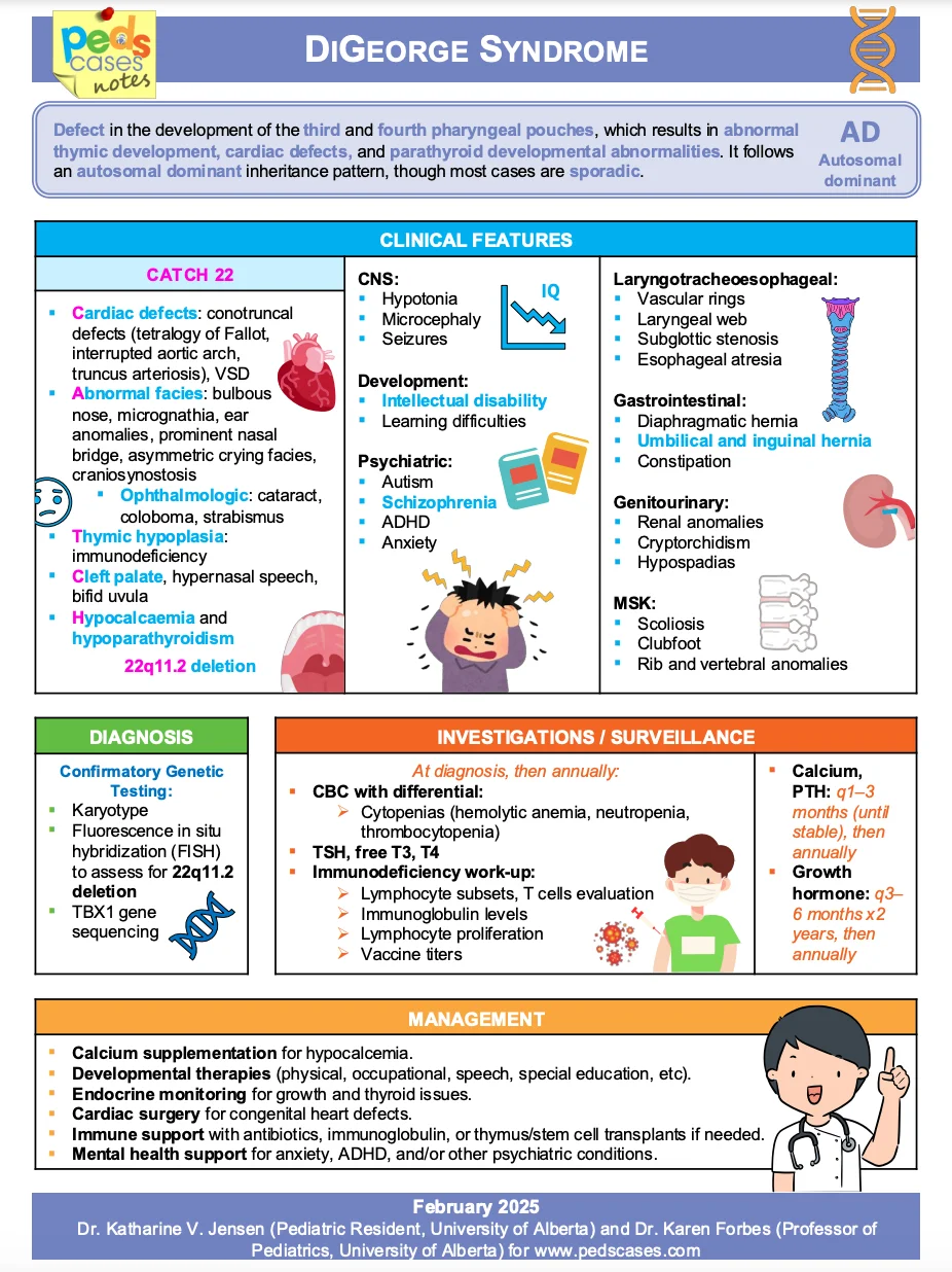
- Heart (75–80%): conotruncal defects, namely tetralogy of Fallot, truncus arteriosus and interrupted aortic arch. A neonate often presents with cyanosis that does not improve with oxygen.
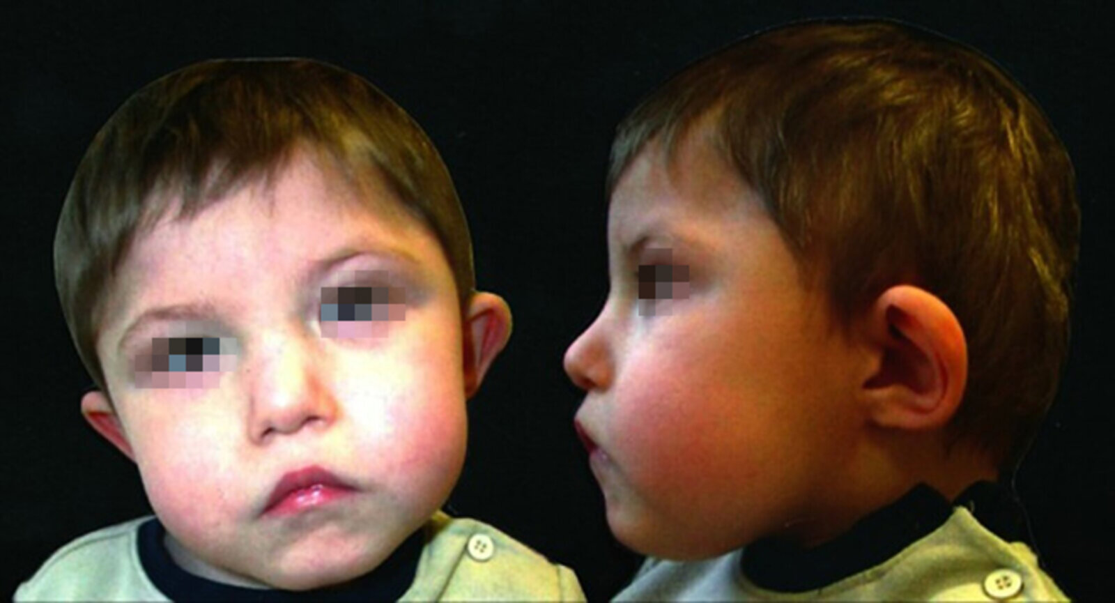
- Face: micrognathia, short philtrum, hypertelorism, and low-set, posteriorly rotated ears; cleft palate or bifid uvula.
- Hypocalcemia: from parathyroid hypoplasia. It causes jitteriness, tetany (Chvostek and Trousseau signs), seizures and arrhythmias in the newborn.
- Immune spectrum:
- Partial DiGeorge (most patients): mildly low T cells with normal B-cell function. Immunity often improves with age.
- Complete DiGeorge (<1%): no thymus and no T cells. B cells are present but cannot make specific antibody, giving SCID-like infections and a risk of GVHD from nonirradiated blood.
- Other: developmental and speech delay, learning disability, later psychiatric illness (eg, schizophrenia), and renal, eye and CNS anomalies.
The face and the heart defect are often what bring the infant to attention.
Diagnosis
- Serum: low calcium with high phosphate and low PTH.
- Immune: lymphopenia on the CBC. Flow cytometry shows low CD3+ T cells with normal B and NK cells.
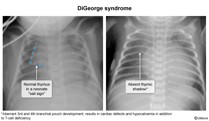
- Imaging: echocardiography for the heart defect; a chest X-ray may show an absent thymic shadow, but a visible thymus does not exclude the diagnosis. Renal ultrasound is also done.
- Most accurate (confirmatory) test: chromosomal microarray (array CGH). FISH for 22q11.2 is the classic alternative.
See the 22q11.2 deletion syndrome summary table for the full CATCH features.
Management
- Hypocalcemia: calcium and calcitriol replacement, lifelong if hypoparathyroidism persists.
- Heart: surgical repair of the conotruncal defect, which may be urgent in a cyanotic neonate.
- Cleft palate: surgery and speech therapy.
- Immune, partial form: antimicrobial prophylaxis if T cells are very low and infections recur; IVIG if antibody production is poor. Give live vaccines only after an immunologist confirms adequate T-cell numbers and function.
- Immune, complete form: isolation, prophylaxis, IVIG and irradiated blood products only. Definitive treatment: thymus tissue implantation, FDA-approved for congenital athymia since 2021. Immune reconstitution takes 6 months or more.
| Important – فكرة سؤال: fixing the immune defect in complete DiGeorge | |
|
The problem is the missing thymus, not the stem cells: the best way to restore T-cell immunity in complete DiGeorge (athymia) is thymus tissue implantation. IVIG, antibiotics and vaccines are only supportive; HSCT is an alternative that works less well because donor T cells, not thymic education, are transferred. |
تذكر |
Severe Combined Immunodeficiency (SCID)
Etiology
- Definition: a group of gene defects that share one result, namely no functional T cells. Cellular and humoral immunity therefore both fail.
- X-linked SCID (most common; historically about half of cases): an IL2RG mutation. It disables the common gamma chain shared by the receptors for IL-2, IL-4, IL-7, IL-9, IL-15 and IL-21, giving no T or NK cells. B cells are present but nonfunctional.
- Adenosine deaminase (ADA) deficiency (autosomal recessive, about 15–20%): toxic adenosine metabolites build up and kill dividing lymphocytes, so T, B and NK cells are all absent.
- Other autosomal recessive forms: JAK3 (behaves like X-linked SCID), RAG1/RAG2 (failed V(D)J recombination) and IL-7 receptor defects.
Clinical features
- Timing: the infant is well at birth (maternal IgG), then develops severe infections within the first few months.
- Highest-yield presentation:
- Overwhelming sepsis and severe recurrent infections with almost any organism
- Chronic lung infection: Pneumocystis pneumonia, CMV pneumonitis, RSV giant-cell pneumonia
- Failure to thrive with chronic diarrhea (eg, persistent rotavirus)
- Examination clues: persistent oral thrush, a candidal diaper rash that does not respond to treatment, and absent tonsils and lymph nodes.
- Live vaccines (rotavirus, BCG, MMR, varicella, oral polio) cause disseminated vaccine-strain infection.
| ملاحظة سريرية | |
|
Never give live attenuated vaccines to a SCID patient; it will be fatal. |
ملاحظة |
Diagnosis
- Screening: newborn TREC assay on the dried blood spot, which shows very low or absent TRECs. Screening has been universal in all US states since 2018.
- CBC: lymphopenia for age (an absolute lymphocyte count usually <2,500/µL in an infant).
- Flow cytometry (key confirmatory step): absent or very low CD3+ T cells (classically autologous T cells <300/µL). The B- and NK-cell pattern points to the gene.
- T-cell function: proliferation to mitogens (PHA) <10% of normal.
- Immunoglobulins: severe hypogammaglobulinemia with no vaccine response. Maternal IgG can mask this in the first months.
- Chest X-ray: absent thymic shadow. Lymph nodes lack germinal centers.
- Most accurate test: gene sequencing (IL2RG, ADA and others).
- Exclude perinatal HIV, which can look identical.
The absent thymus on chest X-ray is shared with complete DiGeorge syndrome.
| Important – فكرة سؤال: absent thymic shadow — SCID or DiGeorge? | |
|
Both give an absent thymic shadow and opportunistic infections. Look for the extras: hypocalcemia, conotruncal heart defect and dysmorphic face → DiGeorge; normal calcium and heart, all immunoglobulins low, chronic diarrhea and thrush → SCID. |
تذكر |
Management
SCID is a pediatric emergency.
- Step 1: Protect immediately. Use protective isolation and withhold all live vaccines, including rotavirus and BCG. Avoid breast milk from a CMV-seropositive mother.
- Step 2: Prevent infection. Give Pneumocystis prophylaxis with TMP-SMX (trimethoprim 150 mg/m²/day orally in 2 divided doses, 3 days a week, from 6 weeks of age), antifungal prophylaxis (eg, fluconazole), monthly IVIG (400–600 mg/kg every 3–4 weeks) to keep IgG normal, and nutritional support.
- Step 3: Definitive treatment. HSCT as early as possible, ideally in the first 3.5 months and before serious infection, when survival exceeds 90%. Gene therapy (eg, for ADA-SCID) is an option when no matched donor exists.
Transfusion rule: any blood product must be irradiated, CMV-negative and leukoreduced. Viable donor T cells in nonirradiated blood cause fatal transfusion-associated graft-versus-host disease.
| فخ امتحاني | |
|
دايماً تذكر إن مريض الـ SCID أو الـ complete DiGeorge ممنوع ياخذ نقل دم عادي، لازم الـ blood products تكون irradiated عشان نمنع الـ Graft-versus-host disease (GVHD). |
ملاحظة |
Untreated SCID is usually fatal in the first year or two of life. The SCID summary table condenses its etiology, inheritance, features and treatment.
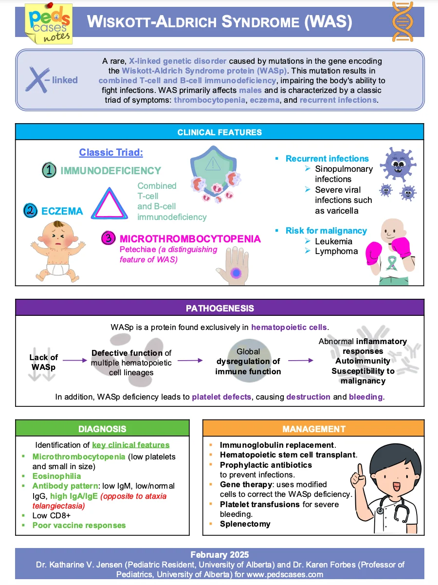
Wiskott-Aldrich Syndrome (WAS)
Etiology
- Genetics: an X-linked recessive mutation of the WAS gene (Xp11.23), so patients are boys.
- Mechanism: the WAS protein, expressed only in blood cells, controls actin cytoskeleton remodeling. Without it, T cells cannot form an immune synapse or migrate normally, and platelets are small and short-lived.
| Mnemonic – جملة تذكرية: eXit (Wiskott-Aldrich) | |
|
جملة تذكرية |
Clinical features
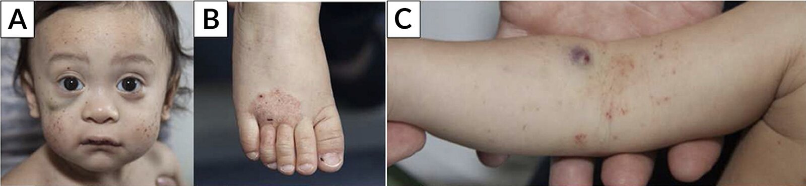
- Bleeding (earliest, from birth): prolonged bleeding after circumcision or from the umbilical stump, petechiae, bloody diarrhea and epistaxis; intracranial hemorrhage in severe cases.
- Eczema: atopic-like, often severe, and frequently infected with S. aureus.
- Infections (worsen with age): recurrent otitis media, sinusitis and pneumonia from encapsulated bacteria (poor antibody to polysaccharides), plus opportunistic infections and severe viral skin infections (HSV, varicella).
- Immune dysregulation: autoimmune cytopenias and vasculitis, and a raised risk of B-cell (often EBV-driven) lymphoma and leukemia, especially in adolescence and early adulthood.
Petechiae next to eczema in a toddler boy is the picture to recognize.
Diagnosis
- CBC and smear (best initial): thrombocytopenia with small platelets (low mean platelet volume), the most consistent finding.
- Immunoglobulins: IgM ↓, IgA ↑ and IgE ↑ (with eosinophilia), IgG normal or low; poor response to polysaccharide vaccines.
- T cells: normal early, then fall in number and lose their mitogen response with age.
- Most accurate (confirmatory) test: WAS gene sequencing, or absent WAS protein on flow cytometry.
| Important – فكرة سؤال: small platelets vs large platelets | |
|
Thrombocytopenia with small platelets (low MPV) in a boy with eczema = Wiskott-Aldrich (a production/cytoskeleton defect). Thrombocytopenia with normal or large platelets after a viral illness and no eczema = ITP (peripheral destruction). The mutated WAS protein regulates the actin cytoskeleton — not DNA repair (AT) or class switching (hyper-IgM). |
تذكر |
An illustrated triad of eczema, purpura and infections brings the presentation, labs and mechanism together.
Management
- Definitive treatment: HSCT from an HLA-matched donor. It is curative and works best when done early.
- Infections: prophylactic antibiotics (TMP-SMX or amoxicillin) and acyclovir as needed; IVIG for a poor antibody response.
- Bleeding: irradiated platelet transfusion for serious bleeding or before surgery; avoid aspirin and other antiplatelet drugs.
- Splenectomy: raises the platelet count when HSCT is not possible, but it deepens the antibody defect and the risk of overwhelming sepsis from encapsulated organisms, so lifelong prophylaxis is needed.
- Eczema: daily emollients and topical corticosteroids.
The Wiskott-Aldrich summary table lists the etiology, features and treatment at a glance.
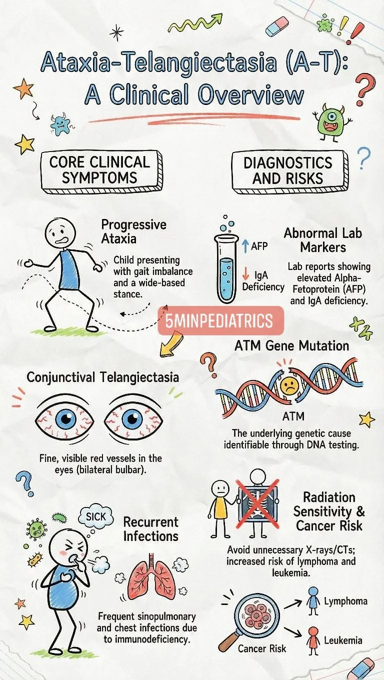
Ataxia-Telangiectasia (AT)
Etiology
- Genetics: an autosomal recessive mutation of ATM (11q22–23).
- Mechanism: ATM kinase senses DNA double-strand breaks and stops the cell cycle so they can be repaired. Without it, breaks accumulate, causing Purkinje cell death, faulty gene rearrangement in developing lymphocytes, genomic instability and hypersensitivity to ionizing radiation.
- Carriers (one mutated ATM copy) have no features of AT but a higher risk of cancer.
Clinical features
- Cerebellar ataxia (first sign): an unsteady gait from the time the child walks, usually clear by age 5. Dysarthria, oculomotor apraxia and loss of motor milestones follow, with severe disability by adolescence.
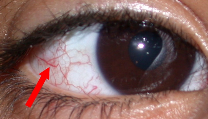
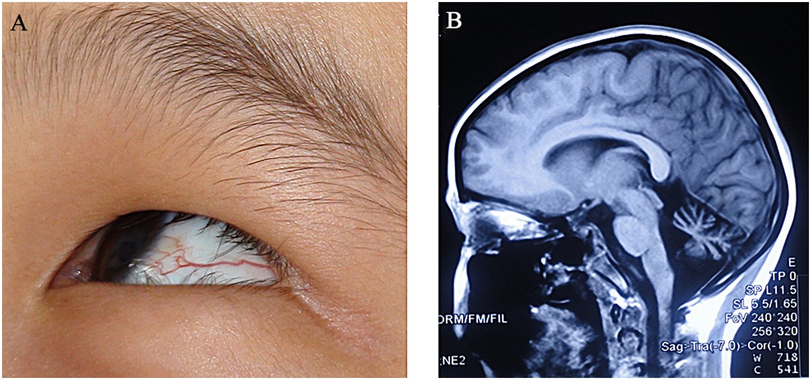
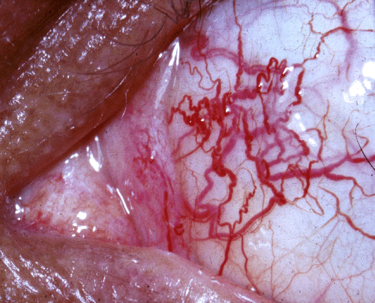
- Telangiectasias (later, after the ataxia): on the bulbar conjunctiva and on sun-exposed skin such as the ears, neck and antecubital fossa.
- Immunodeficiency: recurrent sinopulmonary infections leading to bronchiectasis; IgA, IgE and IgG2 are low while IgM is normal; T-cell lymphopenia with loss of delayed-type hypersensitivity.
- Malignancy: markedly increased risk, mainly lymphoid leukemia and lymphoma.
- Endocrine: insulin resistance and diabetes, poor growth and delayed puberty.
- Course: the neurologic disability outweighs the immune defect. Chronic lung disease with respiratory failure and cancer are the leading causes of death.
The eye finding is often what finally makes the diagnosis.
Diagnosis
- Best initial test: serum alpha-fetoprotein, raised in more than 90% of patients and the most consistent laboratory abnormality.
- Classic picture in a child over 5: ataxia, telangiectasia and a high AFP. Before age 5 the telangiectasias may not have appeared yet, so consider AT in any young child with ataxia and sinopulmonary infections.
- Supporting findings: lymphopenia and low IgA, IgE and IgG2. Low TRECs may appear on newborn screening. Brain MRI is normal early and shows cerebellar atrophy later.
- Most accurate (confirmatory) test: ATM gene sequencing.
- Differential: Friedreich ataxia starts in adolescence with cardiomyopathy and areflexia but no telangiectasia or immune defect.
Management
- Supportive: no treatment halts the neurodegeneration.
- Infections: prophylactic antibiotics; IVIG for hypogammaglobulinemia or a poor antibody response; inactivated vaccines only, no live vaccines.
- Radiation: avoid X-rays and CT whenever possible and prefer ultrasound or MRI. If cancer develops, radiotherapy and chemotherapy must be modified.
- Neurology and nutrition: physical, occupational and speech therapy; a gastrostomy if swallowing is unsafe; screening for diabetes and cancer.
- HSCT: corrects only the immune defect, not the ataxia, and is rarely used.
| فخ امتحاني | |
|
A patient with Ataxia Telangiectasia has a defect in DNA repair, so be cautious about ordering CT scans or any ionizing radiation because of the markedly increased risk of malignancy. |
تذكر |
Key Points for Exams – نقاط مهمة للامتحانات
- T-cell and combined defects present before 4–6 months with failure to thrive, chronic diarrhea, thrush and Pneumocystis pneumonia. B-cell defects present later with encapsulated bacteria.
- Because B cells need T-cell help, a severe T-cell defect lowers antibodies even when B-cell numbers are normal.
- DiGeorge = CATCH 22: 22q11.2 deletion affecting the 3rd/4th pharyngeal pouches, with conotruncal defects (tetralogy of Fallot, truncus arteriosus, interrupted aortic arch), hypocalcemia and thymic hypoplasia.
- The neonate with DiGeorge is most at risk from hypocalcemic seizures. Chromosomal microarray (or FISH) confirms the diagnosis.
- The best way to correct the immune defect in complete DiGeorge is thymus tissue implantation.
- SCID: the most common form is X-linked (IL2RG, common gamma chain); next is autosomal recessive ADA deficiency, the classic target of gene therapy.
- SCID is detected by low TRECs on newborn screening and confirmed by flow cytometry (absent CD3+ T cells). The cure is early HSCT.
- An absent thymic shadow occurs in both SCID and DiGeorge. Hypocalcemia and a heart defect point to DiGeorge.
- Never live vaccines (rotavirus, BCG, MMR, varicella, oral polio) and only irradiated blood in SCID and complete DiGeorge, to prevent transfusion-associated GVHD.
- Wiskott-Aldrich (eXit): X-linked eczema, immunodeficiency and thrombocytopenia with small platelets. Classic case: bleeding after circumcision. Labs: IgM ↓, IgA and IgE ↑.
- WAS protein regulates the actin cytoskeleton. HSCT is curative, and lymphoma risk is high.
- Ataxia-telangiectasia: autosomal recessive ATM DNA-repair defect with ataxia, then telangiectasia, sinopulmonary infections, high AFP, and low IgA, IgE and IgG2.
- In AT, avoid ionizing radiation because of the high risk of leukemia and lymphoma. Death is usually from lung disease or cancer.
احصل على التجربة الكاملة
اشترك للوصول لفيديوهات الشرح التفصيلي والبطاقات التعليمية التفاعلية وأسئلة الممارسة مع تتبع التقدم.