شرح المدرسين

Summary
The esophagus is a 25–30 cm muscular conduit extending from the pharynx (lower border of the cricoid cartilage, C6) to the gastric cardia (T11), transporting the bolus by coordinated peristalsis through the neck, thorax, and abdomen before piercing the diaphragm at the T10 esophageal hiatus. It is a segmentally supplied tube with a rich submucosal vascular and lymphatic plexus and — critically — no serosa along its thoracic course.
The diaphragm is a dome-shaped musculotendinous partition separating the thoracic and abdominal cavities. It is the principal muscle of respiration and a key contributor to gastroesophageal junction (GEJ) competence through the pinch-cock action of the right crus. Command of these relationships underpins the four operations built on this anatomy: esophagectomy, hiatal hernia repair, fundoplication, and antireflux surgery.
Embryology of the Esophagus and Diaphragm
Esophagus
The esophagus arises from the foregut. In the 4th week a respiratory (laryngotracheal) diverticulum buds ventrally, and the tracheoesophageal septum partitions the primitive foregut into a ventral respiratory tract and a dorsal esophagus that elongates as the heart and lungs descend.
Muscle differentiation follows a cranio-caudal rule: the upper third is skeletal muscle derived from branchial (pharyngeal) arch mesenchyme, whereas the lower two-thirds is smooth muscle from splanchnic mesoderm. Failure of foregut septation produces the clinically important trio of esophageal atresia, tracheoesophageal fistula, and congenital esophageal stenosis (esophageal duplication cyst is the rarer fourth).
Diaphragm
The diaphragm is assembled from four embryologic components, each contributing a distinct adult region:
| Embryologic origins of the diaphragm | |
| Embryologic component | Adult diaphragm derivative |
| Septum transversum | Central tendon |
| Pleuroperitoneal membranes | Posterolateral (dorsolateral) diaphragm |
| Dorsal mesentery of the esophagus | Crura of the diaphragm |
| Muscular ingrowth from the body wall | Peripheral muscular rim |
| Mnemonic – جملة تذكرية: Diaphragm embryology | |
|
"Several Parts Definitely Make the diaphragm" → Septum transversum, Pleuroperitoneal membranes, Dorsal mesentery of esophagus, Muscular body wall ingrowth. |
جملة تذكرية |
Defects in this fusion produce congenital diaphragmatic hernias. The congenital diaphragmatic hernia reference traces these lesions back to failure of the pleuroperitoneal fold to close the pericardioperitoneal canal.
Gross Anatomy and Segmental Relations
The adult esophagus measures 25–30 cm (8–10 cm in the newborn) and runs from the lower border of the cricoid cartilage (C6) to the gastric cardia (T11). It is conventionally divided into cervical, thoracic, and abdominal segments, each defined by its own relations and its own operative hazards. Rather than memorise scattered lists, anchor the three most surgically consequential relations of the thoracic segment — the recurrent laryngeal nerves, the thoracic duct, and the left atrium — and read the rest off the master table below.
| The three esophageal segments – length, extent, and key relations | |||
|---|---|---|---|
| Feature | Cervical | Thoracic | Abdominal |
| Length | ~5 cm | 18–20 cm | 2–4 cm |
| Extent | C6 (cricoid) → thoracic inlet | Thoracic inlet → T10 hiatus | T10 hiatus → gastric cardia (T11) |
| Anterior relation | Trachea | Left main bronchus, left atrium & pericardium | Left lobe of liver, anterior vagal trunk |
| Posterior relation | Prevertebral fascia & vertebrae | Thoracic duct, descending aorta | Right crus, posterior vagal trunk |
| Lateral relation | Carotid sheath, thyroid, recurrent laryngeal nerves | Azygos vein (right); aortic arch (left) | Retroperitoneal |
| Outer coat | Adventitia | Adventitia – NO serosa | Serosa (short segment only) |
| Chief surgical hazard | Recurrent laryngeal nerve injury | Thoracic duct injury → chylothorax | Vagal trunk injury during fundoplication |
The anterior relation of the mid-thoracic esophagus to the left atrium explains why a grossly dilated left atrium (e.g., severe mitral stenosis) can indent the esophagus and produce dysphagia, and why a retrocardiac barium column is a useful surrogate for atrial size.
| Note – ملاحظة: cervical esophagus | |
|
The recurrent laryngeal nerves lie in the tracheoesophageal grooves flanking the cervical esophagus and are at risk during cervical esophagectomy, thyroid surgery, and Zenker diverticulum repair — injury produces hoarseness and impaired airway protection. |
ملاحظة |
Normal Esophageal Constrictions
Four physiologic constrictions narrow the esophageal lumen at predictable levels. They are the highest-yield anatomical landmarks of the whole tube because they dictate where pathology and iatrogenic injury concentrate. Learn each by its vertebral level and its endoscopic depth from the incisor teeth.
| The four normal esophageal constrictions | |||
|---|---|---|---|
| Constriction | Vertebral level | Distance from incisors | Anatomical cause |
| Upper esophageal sphincter | C6 | 15 cm | Cricopharyngeus muscle (UES) |
| Aortic arch | T4 | 25 cm | Crossing of the aortic arch |
| Left main bronchus | T5 | 27 cm | Indentation by left main bronchus |
| Diaphragmatic hiatus | T10 | 40 cm | Passage through the diaphragm |
| Mnemonic – جملة تذكرية: constriction distances | |
Endoscopic depths from the incisors climb 15 → 25 → 27 → 40 cm for the Cricopharyngeus, Aortic arch, Bronchus (left main), and Diaphragm — remember "CABD" as you advance the scope. |
جملة تذكرية |
| Important – فكرة سؤال | |
The four constrictions are the sites where a swallowed foreign body impacts, where caustic ingestion causes the deepest burns, and where strictures and instrumentation perforations cluster. The narrowest and most proximal — the cricopharyngeus (UES) — is the single most common site of both foreign-body lodgement and iatrogenic perforation during rigid endoscopy. |
تذكر |
Histology of the Esophagus
The esophageal wall follows the standard gut plan with two clinically decisive modifications — a protective squamous lining and the absence of serosa along the thoracic segment.
- Mucosa — non-keratinized stratified squamous epithelium (built for mechanical abrasion), lamina propria, and muscularis mucosae. The abrupt change to gastric columnar epithelium is the endoscopic Z-line.
- Submucosa — houses the mucous esophageal glands, the vascular and lymphatic plexuses, and the Meissner (submucosal) plexus.
- Muscularis propria — skeletal muscle in the upper third, mixed in the middle third, and smooth muscle in the lower third; the Auerbach (myenteric) plexus lies between its circular and longitudinal layers.
- Adventitia — loose connective tissue over the thoracic esophagus; only the short abdominal segment acquires a true serosa.
| Note – ملاحظة: the two nerve plexuses | |
|
Meissner (submucosal) plexus sits in the submucosa and governs secretion and local blood flow; Auerbach (myenteric) plexus lies between the circular and longitudinal layers of the muscularis propria and drives peristalsis. Their degeneration underlies achalasia. |
ملاحظة |
The surgical importance of the missing serosa cannot be overstated: it removes the tough outer barrier that normally contains tumour and limits leakage, so malignancy extends locally with ease, anastomoses are prone to breakdown, and any perforation vents directly into the mediastinum.
Gastroesophageal Junction and the Antireflux Barrier
The gastroesophageal junction (GEJ) is the transition from esophageal squamous mucosa to gastric columnar mucosa — the endoscopic Z-line. Continence here is not the work of a single sphincter but of an overlapping antireflux barrier, and every antireflux operation is an attempt to reconstruct its components:
- Lower esophageal sphincter (LES) — intrinsic smooth-muscle high-pressure zone.
- Intra-abdominal esophageal segment — positive abdominal pressure keeps it collapsed.
- Diaphragmatic crura — the right crus provides an external "pinch-cock" sphincter.
- Angle of His — the acute esophagogastric angle acts as a flap valve.
- Phrenoesophageal ligament and the gastric fundus — anchor and buttress the junction.
Here is the anatomy that these mechanisms defend, contrasted with the two hernias that defeat them:
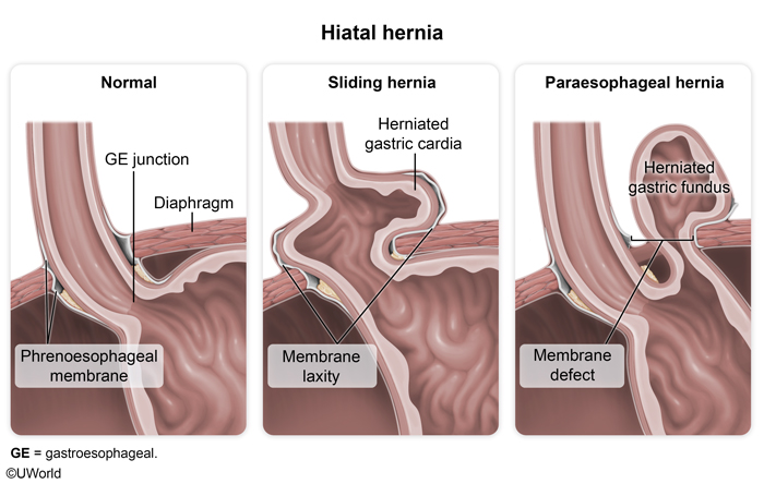
Failure of these mechanisms predisposes to gastroesophageal reflux disease (GERD) and hiatal hernia. The GERD pathophysiology summary links hiatal herniation and LES dysfunction to the reflux phenotype.
| Important – فكرة سؤال | |
The lower esophageal sphincter (LES) is a physiologic high-pressure zone, not a discrete anatomic ring — dissection reveals no thickened muscle. Its resting tone (≈ 10–30 mmHg) is generated by intrinsic smooth-muscle tone plus the external pinch-cock of the right crus, so loss of the intra-abdominal segment (as in a sliding hiatal hernia) uncouples the two and precipitates reflux. |
تذكر |
Arterial and Venous Supply
The esophagus has a segmental (three-tier) blood supply — each segment is fed by whichever regional artery is closest — yet a rich submucosal vascular plexus links these territories. Preserving that plexus during mobilisation is the single most important manoeuvre for avoiding ischaemic anastomotic complications after esophagectomy.
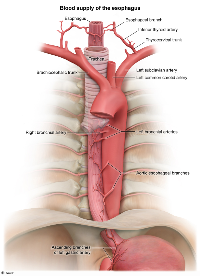
| Segmental arterial supply and venous drainage of the esophagus | ||
|---|---|---|
| Segment | Arterial supply | Venous drainage |
| Cervical | Inferior thyroid artery (from thyrocervical trunk) | Inferior thyroid veins → brachiocephalic veins |
| Thoracic | Esophageal branches of the thoracic aorta + bronchial arteries | Azygos & hemiazygos system → SVC |
| Abdominal | Left gastric artery + left inferior phrenic artery | Left gastric (coronary) vein → portal vein |
This three-segment arterial map is a classic Step 1 vignette: a Mallory-Weiss tear at the GEJ bleeds from the distal territory supplied by the left gastric artery, whereas mid-esophageal lesions bleed from direct aortic branches and cervical lesions from the inferior thyroid artery.
Venous drainage mirrors the arteries but carries a unique clinical twist at its lower end, where portal and systemic systems meet:
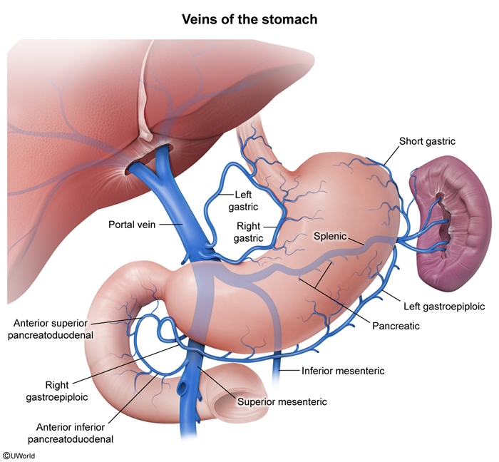
The portosystemic anastomoses reference maps this left-gastric-to-azygos connection alongside the rectal and periumbilical shunts that decompress a hypertensive portal system.
Lymphatic Drainage and Innervation
Lymphatic drainage
Drainage broadly follows the segments, but the defining feature is a dense network of longitudinal submucosal lymphatics that allows tumour to travel far along the wall before it drains to a regional node.
| Lymphatic drainage of the esophagus by level | |
|---|---|
| Segment | Primary nodal station |
| Cervical | Deep cervical nodes |
| Upper thoracic | Paratracheal nodes |
| Middle thoracic | Mediastinal (tracheobronchial) nodes |
| Lower thoracic / distal | Celiac & left gastric nodes |
| Important – فكرة سؤال | |
Esophageal lymphatics run longitudinally within the submucosa, so a small primary tumour can seed distant nodal stations and produce skip metastases far from the visible lesion. This is why oncologic esophagectomy demands an extended (two- or three-field) lymphadenectomy rather than local excision. |
تذكر |
Innervation
Parasympathetic (vagal) fibres form the two vagal trunks on the distal esophagus and drive peristalsis, LES relaxation, and glandular secretion. Sympathetic fibres from the T1–T10 chain mediate vasoconstriction, reduced motility, and visceral pain transmission.
| Mnemonic – جملة تذكرية: vagal trunks | |
"LARP" → Left vagus → Anterior trunk; Right vagus → Posterior trunk. The 90° clockwise rotation of the embryonic foregut carries the left nerve to the front and the right nerve to the back of the distal esophagus. |
جملة تذكرية |
Surgical Anatomy of the Diaphragm
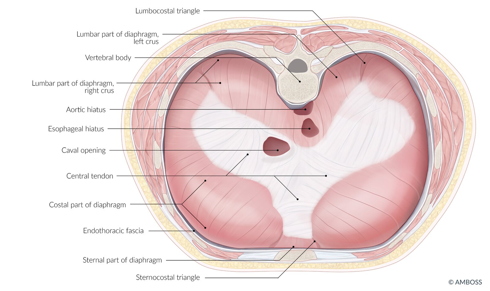
The diaphragm is a dome-shaped musculotendinous sheet with a central tendon and a peripheral muscular rim that takes origin from three attachments: sternal (posterior xiphoid), costal (inner surfaces of ribs 7–12), and lumbar (the crura and arcuate ligaments).
Crura
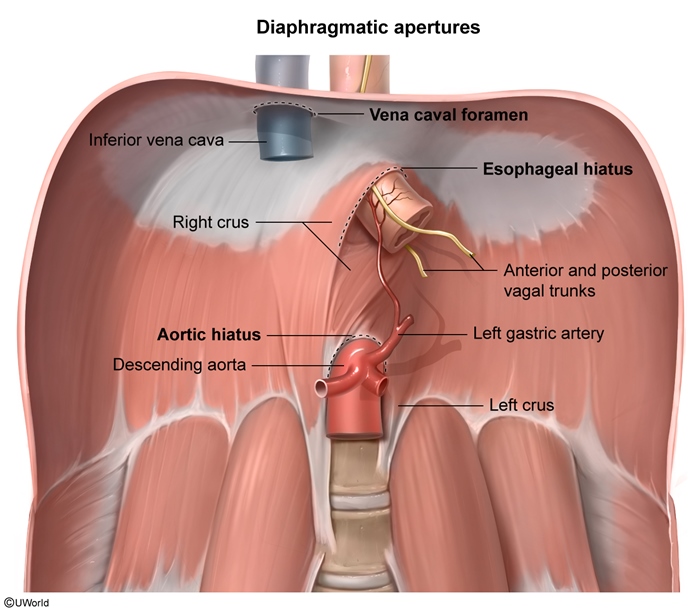
The right crus arises from the bodies of L1–L3, is longer and stronger than the left, and splits to form the esophageal hiatus, slinging around the distal esophagus. The left crus arises from L1–L2 and is shorter. This right-crus sling is the anatomical basis of the diaphragmatic "pinch-cock" antireflux mechanism.
Diaphragmatic apertures
| The three major diaphragmatic apertures | ||
|---|---|---|
| Opening | Vertebral level | Principal contents |
| Caval opening | T8 | Inferior vena cava + right phrenic nerve branches |
| Esophageal hiatus | T10 | Esophagus, anterior & posterior vagal trunks, esophageal branches of left gastric vessels, lymphatics |
| Aortic hiatus | T12 | Aorta, thoracic duct, azygos vein |
| Mnemonic – جملة تذكرية: hiatus levels | |
|
"I 8 (ate) 10 EGGs AT 12" → at T8 the IVC; at T10 the (o)Esophagus + vaGi; at T12 the Aorta, Thoracic duct, and aZygos vein. Note the number of contents climbs 1 → many → 3 as you descend. |
جملة تذكرية |
Phrenoesophageal ligament, angle of His, and phrenic nerve
The phrenoesophageal ligament is a fibroelastic membrane anchoring the distal esophagus to the hiatus; it maintains the intra-abdominal esophageal segment and preserves the antireflux barrier — its weakening is a prime contributor to hiatal hernia. The angle of His, the acute angle between esophagus and gastric fundus, acts as a flap valve, and its loss predisposes to GERD.
The phrenic nerves supply the sole motor innervation to the diaphragm — remember "C3, 4, 5 keep the diaphragm alive." The right phrenic travels with the IVC (through the caval opening), while the left pierces the muscular diaphragm separately. Injury produces hemidiaphragm paralysis with an elevated hemidiaphragm and postoperative respiratory compromise.
Applied Surgical Anatomy and Clinical Correlations
Every relation covered so far earns its place because it changes an operation. The table below is the consolidated "why it matters" list — each structure paired with the complication that follows if it is misjudged.
| Applied surgical anatomy – structure and its clinical consequence | |
|---|---|
| Structure | Clinical importance |
| Recurrent laryngeal nerve | Injury → hoarseness and aspiration |
| Thoracic duct | Injury → chylothorax |
| Left gastric artery | Principal arterial supply to the distal esophagus |
| Right crus | Forms the hiatus; major external antireflux mechanism |
| Vagal trunks | Injury → gastroparesis / delayed gastric emptying |
| Phrenoesophageal ligament | Failure → hiatal hernia |
| Distal submucosal lymphatics | Early longitudinal spread of esophageal carcinoma |
| Absent thoracic serosa | Local tumour extension & mediastinal contamination after perforation |
Hiatal hernia — the anatomy that fails
Laxity or defect of the phrenoesophageal membrane at the hiatus produces the spectrum of hiatal hernias, from the common sliding type to the dangerous paraesophageal type:
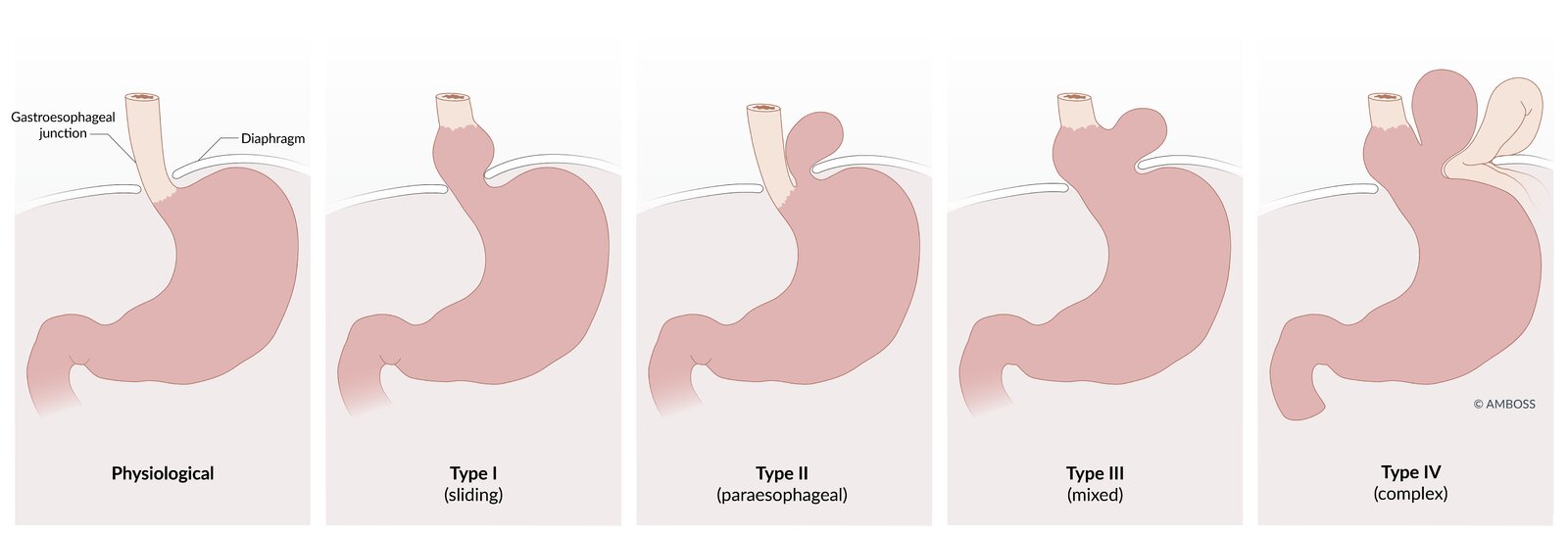
| Sliding vs paraesophageal hiatal hernia | ||
|---|---|---|
| Feature | Sliding (Type I) | Paraesophageal (Type II) |
| Frequency | ~95% – most common | Uncommon |
| Mechanism | Phrenoesophageal membrane laxity | Localised membrane defect |
| GEJ position | Migrates above the diaphragm | Stays below; fundus herniates alongside |
| Dominant risk | GERD | Incarceration, strangulation, volvulus |
| Typical management | Medical (PPI) ± fundoplication | Surgical repair (risk of strangulation) |
Key operative sequences
During esophagectomy, the structures to identify and protect or manage are the recurrent laryngeal nerves, thoracic duct, azygos vein, vagus nerves, left gastric artery, inferior pulmonary veins, aorta, trachea, main bronchi, and pericardium.
Hiatal hernia repair / fundoplication proceeds in a defined order:
- Step 1: Complete circumferential mobilisation of the distal esophagus into the mediastinum to restore an adequate intra-abdominal esophageal segment.
- Step 2: Identify and preserve the anterior and posterior vagal trunks throughout the dissection.
- Step 3: Reduce the hernia sac and expose the right and left crura.
- Step 4: Approximate the crura posteriorly (crural repair) without excessive tension to recalibrate the hiatus.
- Step 5: Construct an appropriate fundoplication (e.g., 360° Nissen) when antireflux control is indicated.
| Important – فكرة سؤال | |
During Nissen fundoplication or hiatal hernia repair, the anterior and posterior vagal trunks lie directly on the distal esophagus and are easily caught in the dissection. Inadvertent vagal injury denervates the pylorus and causes gastroparesis with delayed gastric emptying, early satiety, and bloating — a classic post-operative exam vignette. Always identify and preserve both trunks before crural approximation. |
تذكر |
Mnemonics
The five memory hooks that carry this lesson through a viva or a written paper, consolidated for last-minute revision:
| Mnemonic – جملة تذكرية: esophagus & diaphragm quick-recall | |
|
جملة تذكرية |
Key Points for Exams – نقاط مهمة للامتحانات
- The esophagus runs from C6 to T11 and measures 25–30 cm, divided into cervical, thoracic, and short abdominal segments.
- The thoracic esophagus lacks a serosa, predisposing to rapid local tumour spread and severe mediastinal contamination after perforation.
- Four physiologic constrictions occur at the UES (C6), aortic arch (T4), left main bronchus (T5), and diaphragmatic hiatus (T10) — the sites of foreign-body impaction, caustic injury, and instrumentation perforation.
- The LES is a physiologic high-pressure zone, supported by the right crus, intra-abdominal esophagus, angle of His, and phrenoesophageal ligament — together the antireflux barrier.
- Arterial supply is segmental (inferior thyroid → thoracic aortic/bronchial → left gastric) but the submucosal plexus must be preserved during mobilisation.
- Longitudinal submucosal lymphatics permit extensive proximal and distal (skip) spread of carcinoma, justifying wide lymphadenectomy.
- The distal esophagus is a portosystemic anastomosis (left gastric ↔ azygos), the anatomic basis of esophageal varices in portal hypertension.
- The diaphragm forms from four components; fusion defects cause Bochdalek (posterolateral) and Morgagni (anteromedial) hernias.
- Apertures: T8 IVC, T10 esophagus + vagal trunks, T12 aorta + thoracic duct + azygos; the esophageal hiatus is formed by the right crus.
- The phrenic nerves (C3–C5) are the sole motor supply to the diaphragm; injury causes hemidiaphragm paralysis and respiratory compromise.
- Vagal trunks follow LARP (Left→Anterior, Right→Posterior); injury during fundoplication causes gastroparesis.
احصل على التجربة الكاملة
اشترك للوصول لفيديوهات الشرح التفصيلي والبطاقات التعليمية التفاعلية وأسئلة الممارسة مع تتبع التقدم.