SUMMARY
Gastroesophageal reflux disease (GERD) is a chronic disorder in which reflux of gastric contents into the esophagus causes troublesome symptoms, mucosal injury, or extraesophageal complications. It reflects failure of a multicomponent antireflux barrier — not simply a weak lower esophageal sphincter (LES) — with the balance tipped toward aggressive factors (acid, pepsin, bile) over protective mechanisms (LES, diaphragmatic crura, esophageal clearance, mucosal defense).
The dominant mechanism in most patients is transient LES relaxations (TLESRs), amplified by obesity, sliding hiatal hernia, delayed gastric emptying, and impaired esophageal clearance. Typical symptoms are heartburn and acid regurgitation; atypical/extraesophageal presentations include chronic cough, hoarseness, and asthma. The feared trajectory is chronic esophagitis → peptic stricture → Barrett esophagus → esophageal adenocarcinoma.
Diagnosis is usually clinical, with a PPI trial as first-line therapy. Upper endoscopy is reserved for alarm features and is the gold standard for mucosal complications; 24-hour ambulatory pH monitoring is the gold standard for confirming pathologic acid reflux. Proton pump inhibitors (PPIs) are the most effective medical treatment, and laparoscopic Nissen fundoplication is the standard antireflux operation for medically refractory disease with normal esophageal motility. For a compact overview of mechanism, manifestations, and initial treatment, see the GERD pathophysiology, manifestations, and initial-treatment overview.
Definition, Epidemiology & Risk Factors
Definition
GERD develops when reflux of gastric contents produces troublesome symptoms and/or mucosal injury because the normal antireflux barrier fails and aggressive factors (acid, pepsin, bile salts, pancreatic enzymes) overwhelm protective mechanisms (LES tone, diaphragmatic crura, esophageal peristalsis, salivary bicarbonate, mucosal defense).
Epidemiology
- One of the most common GI disorders worldwide, affecting 10–20% of adults in Western populations.
- Incidence is rising, driven largely by obesity and aging; prevalence is similar in men and women.
- Barrett esophagus and adenocarcinoma cluster in white males > 50 years.
Risk factors (highest-yield)
Truncated to the most commonly tested contributors: central obesity, sliding hiatal hernia, pregnancy, and smoking/alcohol. Scleroderma is the classic connective-tissue cause (LES smooth-muscle atrophy and fibrosis). Among drugs, the ones that lower LES tone and are repeatedly tested are calcium-channel blockers, nitrates, and anticholinergics.
| GERD risk factors – ("OH PS") | |
|
O – Obesity (especially central) Drugs that relax the LES = "CAN": Calcium-channel blockers, Anticholinergics, Nitrates. |
جملة تذكرية |
Pathophysiology & The Antireflux Barrier
GERD results from failure of a multicomponent antireflux barrier rather than a defect in the LES alone. When several of these components fail together, reflux frequency and duration rise and the esophagus is repeatedly bathed in acid.
| Antireflux Barrier Components | |
|---|---|
| Component | Function |
| Lower esophageal sphincter (LES) | Primary barrier — a physiologic high-pressure zone (resting pressure 10–30 mmHg) |
| Diaphragmatic crura | External sphincter — the "pinch-cock" mechanism |
| Intra-abdominal esophagus | Positive intra-abdominal pressure compresses the LES |
| Angle of His | Flap-valve mechanism at the GE junction |
| Phrenoesophageal ligament | Anchors the GEJ below the diaphragm |
| Peristalsis + salivary bicarbonate | Clear and neutralize refluxed acid |
| Normal gastric emptying | Reduces gastric volume and intragastric pressure |
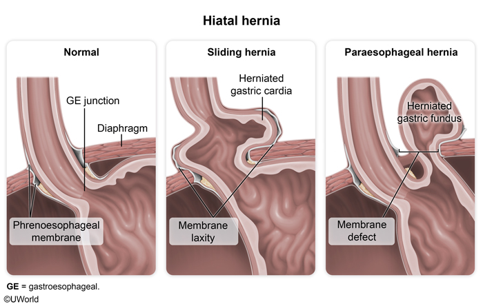
The GE junction anatomy — and how it is distorted by a hiatal hernia — is best appreciated visually:
Why the LES fails
1. Transient LES relaxations (TLESRs) — the most common mechanism. These are relaxations not triggered by swallowing; they are provoked by gastric fundus distension, are physiologic during belching, and cause reflux when they become excessively frequent.
2. Hypotensive LES — inadequate resting pressure from obesity, smoking, scleroderma, or LES-relaxing drugs.
| ملاحظة سريرية – Clinical Note | |
|
ملاحظة سريرية: معظم مرضى الـ GERD الـ LES عندهم ضغط طبيعي — المشكلة الأساسية هي الـ TLESRs، يعني ارتخاءات مؤقتة للـ LES مش مرتبطة بالبلع وبتصير لما يتمدد قاع المعدة (gastric fundus). فلا تفترض دايماً إنه لازم يكون في hypotensive LES. |
ملاحظة |
Hiatal hernia and reflux
A sliding (Type I) hiatal hernia is strongly linked to GERD: the GEJ migrates above the diaphragm, separating the LES from the crura, lowering LES pressure, increasing TLESRs, and creating a supradiaphragmatic acid pocket with impaired clearance. Paraesophageal hernias cause trouble mainly through mechanical complications rather than reflux.
| Sliding vs Paraesophageal Hiatal Hernia | ||
|---|---|---|
| Feature | Sliding hernia (Type I) | Paraesophageal hernia (Types II–IV) |
| Frequency | Most common (>90%) | Uncommon |
| Mechanism | GEJ migrates above the diaphragm → LES separated from crura | GEJ stays fixed; fundus herniates alongside the esophagus |
| Relation to GERD | Strongly associated with reflux | Less associated with reflux |
| Main danger | ↓ LES pressure, ↑ TLESRs, impaired acid clearance | Mechanical obstruction, gastric volvulus, ischemia |
The injury cascade
LES dysfunction + hiatal hernia + delayed gastric emptying + impaired esophageal clearance + acid/bile reflux → repeated mucosal injury → esophagitis → ulceration → stricture / Barrett esophagus → adenocarcinoma.
Clinical Features & Diagnosis
Typical symptoms
Heartburn and acid regurgitation are the hallmark features, classically worse after meals and when supine. Also seen: water brash (reflex salivation), and — as later/complication features — dysphagia and odynophagia.
Extraesophageal symptoms (highest-yield)
Reflux can present without heartburn ("silent GERD"). The most commonly tested atypical presentations are chronic cough, hoarseness/laryngitis, and asthma; dental enamel erosion is a classic exam clue.
Alarm symptoms
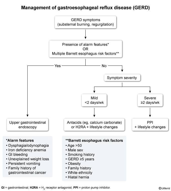
Any alarm feature mandates prompt upper GI endoscopy to exclude malignancy, stricture, or ulceration — do not simply escalate acid suppression.
| Alarm symptoms → endoscopy ("ALARMS") | |
|
A – Anemia (iron deficiency) Any alarm feature → upper GI endoscopy to exclude malignancy, stricture, or ulceration. |
جملة تذكرية |
Diagnostic work-up (by modality)
GERD is largely a clinical diagnosis; a symptomatic response to a PPI trial supports it. Objective testing is reserved for alarm features, atypical/refractory symptoms, or preoperative evaluation:
- Upper endoscopy (EGD): gold standard for mucosal complications — esophagitis, Barrett esophagus, stricture, and cancer; enables biopsy.
- Ambulatory 24-hour pH monitoring: gold standard for confirming pathologic acid reflux; used when endoscopy is normal and always before antireflux surgery.
- High-resolution manometry: assesses LES pressure and esophageal motility; mandatory before surgery to exclude achalasia and major motility disorders.
- Barium swallow: defines anatomy — hiatal hernia, stricture, and short esophagus.
Complications
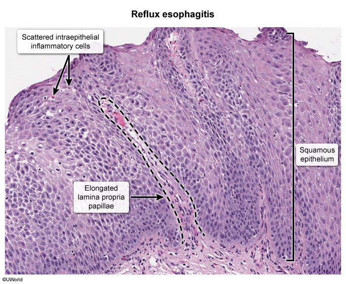
1. Reflux esophagitis
Acid-induced mucosal inflammation presenting with heartburn, chest pain, and dysphagia; ulceration and bleeding may follow. The classic histologic triad is elongated lamina propria papillae, intraepithelial eosinophils/neutrophils, and basal-zone hyperplasia.
2. Peptic stricture
Chronic inflammation → fibrosis → luminal narrowing, presenting with progressive dysphagia to solids. On barium the narrowing is concentric and symmetric (contrast with the asymmetric, irregular narrowing of cancer). Treat with endoscopic dilation + PPI therapy.
3. Barrett esophagus (metaplastic complication)
Replacement of normal stratified squamous epithelium by intestinal-type columnar epithelium with goblet cells — the principal premalignant lesion of chronic GERD. Diagnosis requires upper endoscopy + biopsy confirming specialized intestinal metaplasia. Risk factors mirror adenocarcinoma: long-standing GERD, male sex, white race, age >50, central obesity, and smoking. Endoscopically it appears as salmon-pink tongues extending above the Z-line.
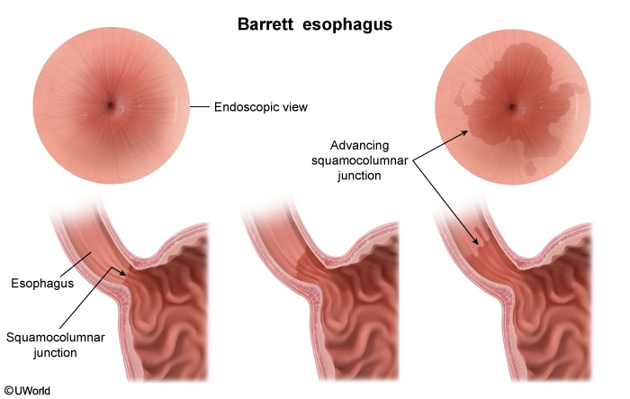
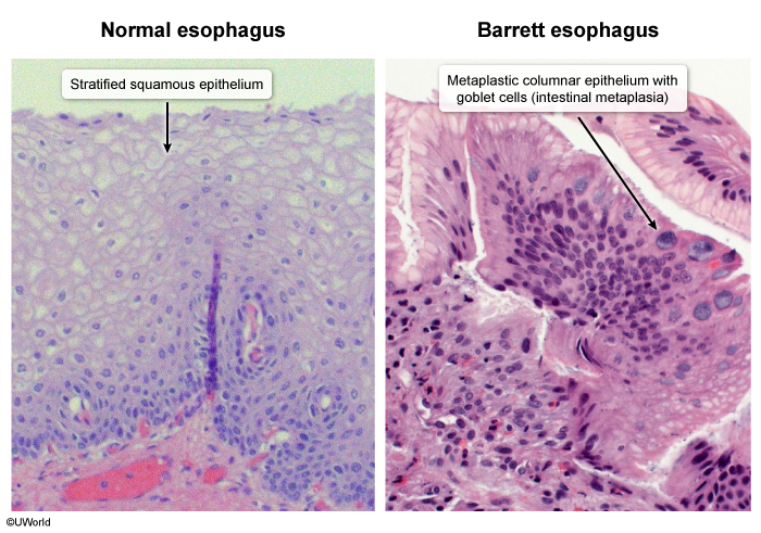
Surveillance intensity is dictated by the highest grade of dysplasia found:
| Barrett Esophagus – Surveillance by Dysplasia Grade | |
| Dysplasia Grade | Histology |
| No dysplasia | Endoscopic surveillance every 3–5 years |
| Low-grade dysplasia | Endoscopic eradication therapy or close surveillance |
| High-grade dysplasia | Endoscopic eradication (radiofrequency ablation ± EMR); esophagectomy in selected cases |
4. Esophageal adenocarcinoma (neoplastic complication)
The metaplasia–dysplasia–carcinoma sequence: GERD → Barrett → low-grade → high-grade dysplasia → invasive adenocarcinoma, most commonly involving the distal esophagus and GEJ. Presents with progressive dysphagia, weight loss, odynophagia, and GI bleeding.
5. Respiratory / extraesophageal complications
Two mechanisms: microaspiration of acid into the airway → inflammation, chronic cough, bronchospasm, and aspiration pneumonia; and a vagal reflex from acid in the distal esophagus → bronchoconstriction. Manifestations include asthma, chronic cough, laryngitis/hoarseness, chronic sinusitis, and dental erosion.
| Important – Question Idea / فكرة سؤال | |
|
Match the symptom to the complication: new-onset odynophagia in chronic GERD → erosive esophagitis / ulcer. Progressive dysphagia to solids only with concentric, symmetric distal narrowing on barium → benign peptic stricture (treat with dilation + PPI). By contrast, asymmetric, irregular narrowing plus weight loss points to adenocarcinoma. Barrett metaplasia itself is typically asymptomatic. |
تذكر |
Management (Medical & Surgical)
Lifestyle modification (first step)
Highest-yield measures: weight loss, smoking cessation, elevating the head of the bed, and avoiding meals within 2–3 hours of sleep plus trigger foods (fatty meals, alcohol, caffeine, chocolate, peppermint) and tight clothing.
Pharmacologic therapy
Proton pump inhibitors (PPIs) — first-line. Examples: omeprazole, pantoprazole, esomeprazole, rabeprazole. They deliver the most effective acid suppression, heal erosive esophagitis, relieve symptoms, and prevent recurrence. They act at the final common pathway of acid secretion — the H⁺/K⁺-ATPase proton pump — which is why they outperform H2 blockers that block only the histamine limb.
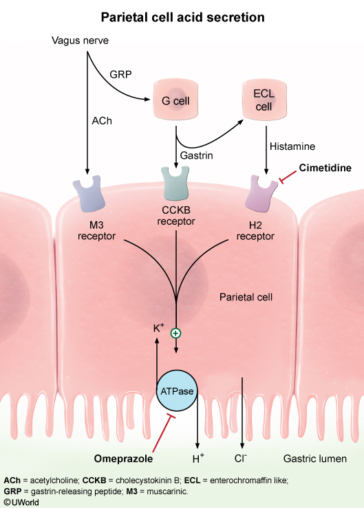
- H2-receptor blockers (famotidine, cimetidine): less effective than PPIs; useful for mild disease or nocturnal symptoms.
- Antacids: rapid symptom relief, no mucosal healing.
- Alginates: form a floating raft over gastric contents to reduce postprandial reflux.
- Prokinetics (metoclopramide): increase gastric emptying and LES tone; limited by adverse effects.
- Baclofen: reduces TLESRs; used in refractory reflux but limited by CNS side effects.
Step-up therapeutic approach
- Step 1: Institute lifestyle modification for typical GERD symptoms; escalate if symptoms persist.
- Step 2: Start a once-daily PPI for 8 weeks and reassess at completion.
- Step 3: If symptoms resolve → continue the lowest effective dose. If they persist → proceed to upper endoscopy ± ambulatory pH monitoring, and consider antireflux surgery if objective GERD is confirmed.
Surgical therapy
Indications: failure of medical therapy, patient preference to avoid lifelong PPIs, severe regurgitation, large hiatal hernia, complications (stricture, recurrent aspiration), and objective evidence of GERD. The ideal candidate has typical reflux symptoms, an excellent PPI response, objectively documented reflux, and normal or correctable esophageal motility.
Preoperative evaluation combines upper endoscopy, high-resolution manometry, and ambulatory pH monitoring (barium swallow and gastric-emptying study in selected patients). The surgical goals are to restore an intra-abdominal esophagus, repair the hiatal defect, and reconstruct the LES without causing obstruction.
The individual antireflux operations, their ideal candidate, and their characteristic complications are consolidated here:
| Procedure | Wrap | Best candidate | Reflux control | Key complications |
| Nissen fundoplication (gold standard) | Complete 360° wrap of the fundus | Normal esophageal motility | Excellent, durable long-term control | Dysphagia, gas-bloat syndrome, inability to belch/vomit |
| Toupet fundoplication | 270° posterior partial wrap | Weak / impaired motility | Slightly higher reflux recurrence than Nissen | Lower dysphagia rate |
| Dor fundoplication | 180–200° anterior partial wrap | Often after Heller myotomy for achalasia | Less effective than Nissen | Least postoperative dysphagia |
| Collis gastroplasty + fundoplication | Stapled tubular lengthening of esophagus + wrap | Shortened esophagus (cannot be reduced) | Creates tension-free intra-abdominal segment | Those of the added fundoplication |
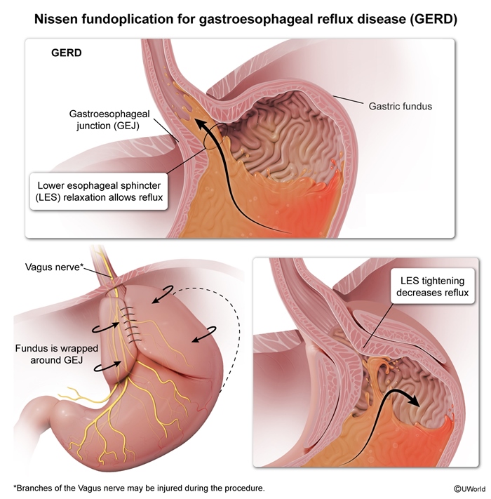
Laparoscopic Nissen fundoplication achieves symptom relief in 85–95% of appropriately selected patients. Early complications include dysphagia, bleeding, and esophageal/gastric perforation; late complications include gas-bloat syndrome, wrap migration, recurrent hiatal hernia, recurrent GERD, and a slipped or disrupted wrap.
| ملاحظة سريرية – Clinical Note | |
|
ملاحظة سريرية: القاعدة البسيطة — Nissen (360°) للمريض اللي الـ esophageal motility عنده طبيعي، وToupet (270° posterior) للمريض اللي عنده weak/impaired motility عشان تقلل خطر الـ postoperative dysphagia. والـ Dor (180° anterior) منعمله عادة بعد Heller myotomy للـ achalasia. |
ملاحظة |
Antireflux surgery in Barrett esophagus
Fundoplication controls reflux, heals esophagitis, and may stabilize Barrett mucosa — but it does not reliably reverse Barrett or eliminate the adenocarcinoma risk, so endoscopic surveillance must continue after a successful operation.
Key Points for Exams
The single table below distills every commonly tested "one-liner" for GERD — mechanism, gold-standard tests, best medical and surgical options, and the two feared complications.
| GERD – High-Yield Summary | |
| Main mechanism of GERD | Transient LES relaxations (TLESRs) |
| Most important antireflux structure | Lower esophageal sphincter (LES) |
| Hiatal hernia associated with GERD | Sliding (Type I) |
| Gold standard for mucosal complications | Upper endoscopy (EGD) |
| Gold standard for objective reflux diagnosis | 24-hour ambulatory pH monitoring |
| Mandatory before antireflux surgery | High-resolution manometry (exclude achalasia) |
| Best medical treatment | Proton pump inhibitors (PPIs) |
| Gold standard surgical procedure | Laparoscopic Nissen fundoplication |
| Best operation for poor esophageal motility | Toupet fundoplication |
| Short esophagus | Collis gastroplasty + fundoplication |
| Premalignant complication | Barrett esophagus |
| Malignant complication | Esophageal adenocarcinoma |
| Key Points for Exams – نقاط مهمة للامتحانات (Schwartz pearls) | |
|
ملاحظة |
احصل على التجربة الكاملة
اشترك للوصول لفيديوهات الشرح التفصيلي والبطاقات التعليمية التفاعلية وأسئلة الممارسة مع تتبع التقدم.