شرح المدرسين

Summary
Congenital anomalies of the upper respiratory tract are structural problems of the larynx and trachea present at birth. They are an important cause of stridor and respiratory distress in newborns and infants. The four main conditions to know are:
- Laryngomalacia — floppy supraglottic cartilage; the most common cause of stridor in infants.
- Subglottic stenosis — narrowing below the vocal cords; the second most common cause of stridor in neonates.
- Vocal cord paralysis — damage to the recurrent laryngeal nerve; weak cry and aspiration risk.
- Laryngeal web — failed recanalization of the larynx during fetal development; abnormal voice and stridor.
Most are diagnosed clinically and confirmed by flexible laryngoscopy. Many mild cases resolve on their own; severe cases need surgery.
Quick orientation to the upper airway
Before going through each condition, it helps to picture where the problem sits in the airway. The type of stridor you hear points to the level of obstruction.
Sagittal anatomy of the upper airway shows the supraglottis (above the cords), the glottis (the cords themselves), and the subglottis (just below the cords) — each is the site of a different congenital anomaly.
| Stridor type — جملة تذكرية | |
|
جملة تذكرية |
Laryngomalacia
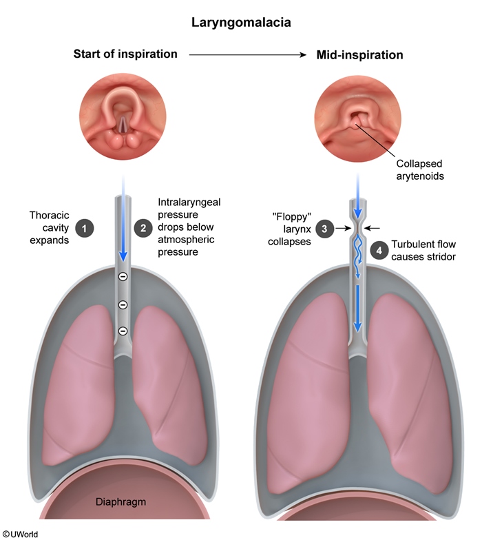
Laryngomalacia is the most common cause of stridor in newborns and infants. The supraglottic cartilage (epiglottis and arytenoids) is not stiff enough yet, so during inspiration the negative pressure sucks these floppy tissues inward and narrows the airway. The result is a noisy, fluttering inspiratory stridor.
Pathophysiology
- Increased laxity of the supraglottic structures.
- On inspiration → negative intralaryngeal pressure → collapse of the floppy supraglottis → turbulent airflow → inspiratory stridor.
The mechanism of inspiratory stridor in laryngomalacia shows how the floppy larynx collapses inward during inspiration.
Clinical presentation
- Inspiratory stridor — may start at birth, usually heard by 2 weeks of age.
- Peaks at 4–8 months.
- Worse with: agitation, crying, feeding, supine position.
- Better with: prone position, neck extension, calm state.
- Severe cases → feeding difficulty, failure to thrive, obstructive hypoxia at night.
Diagnosis
- Mostly a clinical diagnosis.
- Confirmed by awake flexible laryngoscopy in moderate/severe cases.
- Classic finding: omega-shaped (Ω) epiglottis and collapse of the supraglottic structures during inspiration.
Management
- Reassurance — most cases resolve spontaneously by 12–24 months (often around 18 months) as the cartilage stiffens with growth.
- Treat associated GERD if present (it can worsen symptoms).
- Supraglottoplasty (trimming the floppy supraglottis) — reserved for severe cases with feeding difficulty, failure to thrive, or significant obstructive hypoxia.
For a side-by-side review with other causes of infant stridor, see the Differential Diagnosis of Stridor in Infants & Children — it lays out laryngomalacia next to croup, vascular ring, hemangioma, and foreign body.
Subglottic Stenosis
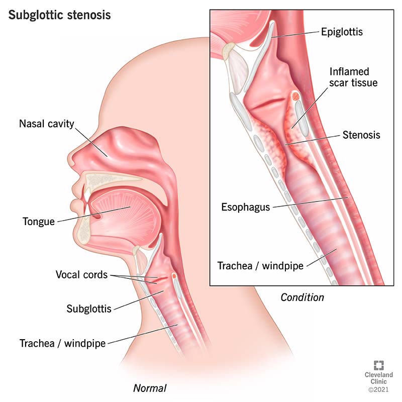
Subglottic stenosis is narrowing of the airway just below the vocal cords (the cricoid region). It is the second most common cause of stridor in neonates. It can be congenital or acquired — and the acquired form is what you see after prolonged intubation.
Pathophysiology
- Congenital: incomplete development of the subglottic area / cricoid cartilage.
- Acquired: scarring after endotracheal intubation — risk rises with longer intubation time. Also after trauma or infection.
- The subglottis is the narrowest part of the infant airway, so even small amounts of swelling or scarring cause big obstruction.
Clinical presentation
- Stridor and respiratory distress in the newborn or first few months of life.
- Stridor is usually biphasic (inspiratory + expiratory) and not affected by position — this helps separate it from laryngomalacia.
- Milder cases → recurrent croup, especially before 12 months of age. Any infant with repeated croup episodes deserves a workup for subglottic stenosis.
Increased incidence in infants with Trisomy 21 (Down syndrome). On a plain neck X-ray, the subglottic area may show fixed narrowing similar to the steeple sign seen in croup, but in stenosis the narrowing is fixed, not transient.
Diagnosis
- Clinical suspicion + direct laryngoscopy or bronchoscopy to visualize and grade the narrowing (gold standard).
- Neck X-ray may suggest the diagnosis but is not definitive.
Management
- Asymptomatic stenosis → observation only.
- Symptomatic → surgical correction (e.g., laryngotracheal reconstruction or cricoid split).
- Some infants need a tracheostomy first to secure the airway until definitive surgery can be performed.
| Note – ملاحظة | |
|
Down syndrome (Trisomy 21) is strongly associated with congenital subglottic stenosis. If a child with Down syndrome has recurrent croup-like episodes, think subglottic stenosis until proven otherwise — and use a smaller endotracheal tube than usual if intubation is needed. |
ملاحظة |
Vocal Cord Paralysis
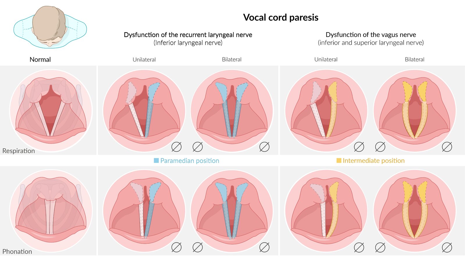
Vocal cord paralysis in children is caused by damage to the recurrent laryngeal nerve (RLN). It can be one-sided (unilateral) or two-sided (bilateral), and the symptoms differ from each other.
Causes
- Neurologic abnormalities: Arnold-Chiari malformation, posterior fossa tumor, hydrocephalus.
- Birth trauma: difficult delivery, neck traction or stretching.
- Iatrogenic: complication of cardiothoracic surgery, thyroid surgery, or tracheoesophageal fistula repair.
- Intubation: pressure injury, most often on the left.
Look at the recurrent laryngeal nerve anatomy to understand why the left side is more vulnerable. The left RLN course around the aortic arch explains the classic exam association.
Clinical presentation
- Unilateral paralysis: weak or hoarse, breathy cry; aspiration risk during feeding; voice quality is "raspy."
- Bilateral paralysis: severe stridor and respiratory distress — both cords sit near the midline and cannot abduct. Often needs urgent airway support.
- Older children → subdued, hoarse voice with limited pitch range.
Diagnosis
- Awake fiberoptic nasopharyngoscopy / flexible laryngoscopy — direct visualization showing absent or impaired vocal cord movement (gold standard).
- Workup for an underlying neurologic cause if no obvious surgical trigger: MRI brain (Arnold-Chiari, posterior fossa lesions, hydrocephalus).
Management
- Unilateral: often improves spontaneously over months; supportive care, feeding therapy to prevent aspiration. Surgical vocal cord medialization (e.g., injection) if persistent.
- Bilateral: secure the airway first — often requires tracheostomy. Definitive vocal cord lateralization procedures can be done later.
- Treat the underlying cause if reversible (e.g., decompression of hydrocephalus).
| Important – فكرة سؤال | |
|
Why is the LEFT recurrent laryngeal nerve injured more often? Because it takes a longer route — it loops under the aortic arch before going back up to the larynx, while the right side simply loops under the right subclavian artery. This is why left-sided vocal cord paralysis is more common after intubation, cardiothoracic surgery, or aortic aneurysm. |
تذكر |
Laryngeal Web
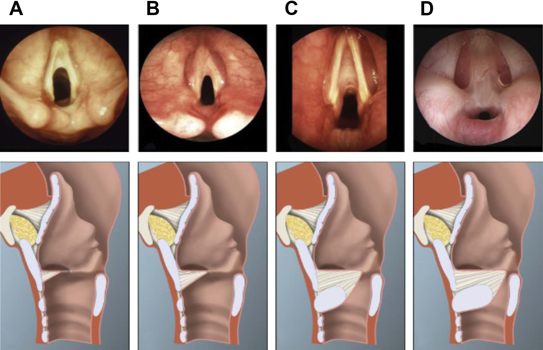
A laryngeal web is a thin membrane of tissue across the laryngeal lumen — usually at the level of the vocal cords (glottic web). It happens because the larynx fails to fully recanalize during fetal development.
Pathophysiology
- During gestation the larynx is a solid tube that normally hollows out (recanalizes). Partial recanalization leaves a web of tissue between the cords.
- Most webs are glottic (at the vocal cords); supraglottic and subglottic webs are less common.
Clinical presentation
- Abnormal voice / weak cry in newborns.
- Stridor and respiratory distress — severity depends on how much of the airway is blocked.
- Severe webs → airway emergency at birth.
Associated syndrome
- Strong association with velocardiofacial syndrome / DiGeorge syndrome (22q11.2 deletion). Always check for cardiac defects, hypocalcemia, and immune problems if a laryngeal web is found.
Diagnosis
- Direct laryngoscopy — visualizes the web (gold standard).
Management
- Thin webs → endoscopic incision or dilation.
- Thick or severe glottic webs → tracheostomy followed by definitive surgical repair (laryngotracheal reconstruction).
| Laryngeal web → تذكر DiGeorge — CATCH-22 | |
|
Laryngeal webs are seen in velocardiofacial syndrome (DiGeorge syndrome, 22q11.2 deletion). Remember the classic CATCH-22 features:
|
جملة تذكرية |
Side-by-side comparison
This table summarizes the four conditions side-by-side. It is the single highest-yield page for exam review.
| Comparison of the four congenital upper airway anomalies | ||||
|---|---|---|---|---|
| Feature | Laryngomalacia | Subglottic stenosis | Vocal cord paralysis | Laryngeal web |
| Site of problem | Supraglottis (above cords) | Subglottis (below cords) | Glottis (vocal cords) | Glottis (between cords) |
| Mechanism | Floppy supraglottic cartilage collapses inward | Congenital or post-intubation narrowing | Recurrent laryngeal nerve damage | Failed recanalization of larynx in utero |
| Type of stridor | Inspiratory; positional (worse supine) | Biphasic; not positional | Variable; weak cry if unilateral | Biphasic if severe; weak cry |
| Onset | Usually by 2 weeks; peaks 4–8 mo | Neonatal or first months of life | Newborn or after surgery/intubation | At birth |
| Classic association | Most common cause of infant stridor | Trisomy 21 (Down syndrome); post-intubation | Arnold-Chiari, cardiothoracic / thyroid surgery | DiGeorge syndrome (22q11.2 deletion) |
| Diagnosis | Flexible laryngoscopy — omega-shaped epiglottis | Direct laryngoscopy / bronchoscopy | Awake fiberoptic laryngoscopy | Direct laryngoscopy |
| Management | Reassurance; supraglottoplasty if severe | Observe if mild; surgery ± tracheostomy | Supportive; tracheostomy if bilateral | Incision / dilation; tracheostomy if severe |
| Prognosis | Resolves by 12–24 months | Depends on severity | Unilateral often improves; bilateral worse | Depends on web thickness |
Key Points for Exams
| Key Points for Exams – نقاط مهمة للامتحانات | |
|
تذكر |
احصل على التجربة الكاملة
اشترك للوصول لفيديوهات الشرح التفصيلي والبطاقات التعليمية التفاعلية وأسئلة الممارسة مع تتبع التقدم.