Summary
Acute ischemic stroke (AIS) is the sudden onset of a focal neurological deficit caused by occlusion of a cerebral artery, resulting in infarction of brain tissue. The single most time-critical distinction is from hemorrhagic stroke — an urgent non-contrast head CT must exclude bleeding before any thrombolytic is given — and from a transient ischemic attack (TIA), in which the deficit resolves without permanent infarction.
Overview
Acute ischemic stroke (AIS) is the sudden onset of a focal neurological deficit caused by occlusion of a cerebral artery, leading to infarction (death) of the brain tissue supplied by that vessel.
Ischemic stroke accounts for roughly 85–87% of all strokes (the remainder being hemorrhagic) and is a leading global cause of death and long-term disability. The core governing principle is "time is brain": an estimated 1.9 million neurons are lost each minute an occlusion persists, so the entire management pathway is built around rapid, time-based decisions to salvage the recoverable penumbra.
- Core concept: focal hypoperfusion → irreversible infarct core surrounded by salvageable, hypoperfused penumbra.
- Goal of acute care: rapidly exclude hemorrhage, then reperfuse eligible patients (thrombolysis ± thrombectomy).
- Mechanistic classification is summarised below using the TOAST framework.
| Ischemic Stroke Subtypes (TOAST Classification) | ||
|---|---|---|
| Category | Subtype | Pathophysiology / Clinical Features |
| Large-vessel | Large-artery atherosclerosis | Thrombotic / artery-to-artery embolism; atheroma of carotid, vertebral, or large intracranial arteries |
| Cardioembolic | Cardioembolism | Atrial fibrillation, MI, valve disease; sudden, maximal deficit; often multiple vascular territories |
| Small-vessel | Lacunar infarct | Lipohyalinosis of penetrating arteries; deep infarcts <15 mm; chronic hypertension and diabetes |
| Other / Cryptogenic | Undetermined or other cause | Dissection, hypercoagulable, vasculitis; younger patients; no cause found despite full workup |
Epidemiology & Etiology
Epidemiology
- Incidence rises sharply with age; most strokes occur after 65 years.
- Slightly more common in men, but women have higher lifetime risk and mortality.
- Leading cause of acquired long-term disability worldwide.
Non-modifiable risk factors
- Advancing age, male sex, family history, prior stroke/TIA, and certain ethnicities.
Modifiable risk factors (the highest-yield exam targets)
- Hypertension — the single most important modifiable factor for both ischemic and hemorrhagic stroke.
- Atrial fibrillation — the classic cause of cardioembolic stroke.
- Diabetes mellitus, dyslipidemia, smoking, obesity, physical inactivity.
- Carotid stenosis, sickle cell disease, and hypercoagulable states.
| Modifiable stroke risk factors — "HEADS" | |
|
جملة تذكرية |
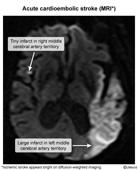
A cardioembolic source (e.g., atrial fibrillation) classically produces sudden, maximal deficits and may seed multiple vascular territories simultaneously, as shown on diffusion imaging.
Pathophysiology
Arterial occlusion causes an abrupt fall in cerebral blood flow (CBF) and triggers the ischemic cascade:
- Energy failure: loss of oxygen/glucose → ATP depletion → failure of the Na⁺/K⁺-ATPase.
- Depolarization & excitotoxicity: membrane depolarizes → excess glutamate release → NMDA-receptor overactivation → intracellular Ca²⁺ influx.
- Cell death: Ca²⁺ activates proteases, lipases, and free radicals → neuronal necrosis and apoptosis.
Core vs. penumbra — the central infarct core (CBF <10 mL/100 g/min) is irreversibly dead, while the surrounding penumbra is hypoperfused but still viable. The penumbra is the target of all reperfusion therapy and is salvageable only within a narrow time window.
| Important – فكرة سؤال | |
A favorite board stem asks for the final common mechanism of neuronal death in ischemia: ATP depletion halts the Na⁺/K⁺-ATPase → membrane depolarization → massive glutamate release → overactivation of NMDA receptors → lethal intracellular Ca²⁺ influx (excitotoxicity). This Ca²⁺ overload activates proteases, lipases, and free-radical pathways that destroy the neuron. |
تذكر |
Mechanism-specific pathology:
- Thrombotic: atherosclerotic plaque rupture at large-artery bifurcations.
- Embolic: cardiac or artery-to-artery emboli lodging distally.
- Lacunar: chronic hypertension/diabetes drive lipohyalinosis and microatheroma of deep penetrating (lenticulostriate) arteries.
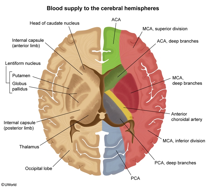
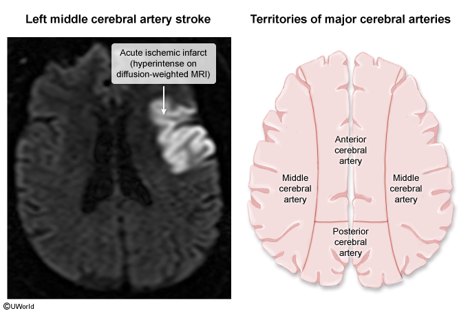
Lacunar disease of the small penetrating lenticulostriate arteries underlies the classic lacunar syndromes. The territory supplied by each major vessel is mapped below.
Clinical Features
The hallmark is the sudden onset of a focal neurological deficit referable to a single vascular territory. The pattern localizes the lesion:
| Anterior vs. Posterior Circulation Stroke Syndromes | |||
|---|---|---|---|
| Feature | ACA | MCA | PCA |
| Motor/sensory loss | Contralateral leg > arm | Contralateral face & arm > leg | Usually spared |
| Cortical signs | Abulia, urinary incontinence | Aphasia (dominant), neglect (non-dominant) | Alexia without agraphia (dominant) |
| Visual | None | Gaze deviation toward lesion | Homonymous hemianopia, macular sparing |
| Classic clue | "Leg weakness" | "Face/arm weakness + speech" | "Isolated vision loss" |
- Posterior/vertebrobasilar: vertigo, diplopia, dysarthria, ataxia, and crossed signs (ipsilateral face, contralateral body).
- Lacunar syndromes: pure motor hemiparesis, pure sensory stroke, ataxic hemiparesis, and dysarthria–clumsy hand — without cortical signs (no aphasia/neglect).
Refer to the lacunar stroke syndromes comparison for the anatomical location mapped to each classic lacunar presentation.
A PCA-territory infarct produces the classic contralateral homonymous hemianopia with macular sparing, reflecting occipital cortex involvement.
Diagnosis
Diagnosis is clinical + emergent imaging, performed in parallel with resuscitation. Group the workup by modality:
- Imaging – first-line: Non-contrast head CT (NCCT) to exclude hemorrhage. Early ischemic signs include the hyperdense MCA sign and loss of grey–white differentiation; the parenchyma is often normal in the first hours.
- Imaging – most sensitive: Diffusion-weighted MRI (DWI) detects restricted diffusion within minutes — the gold standard for confirming acute infarction.
- Vascular imaging: CT angiography identifies large-vessel occlusion (LVO); CT perfusion defines core vs. salvageable penumbra for extended-window thrombectomy.
- Labs: fingerstick glucose (mandatory), CBC, coagulation panel (PT/INR, aPTT), troponin, and 12-lead ECG (detect atrial fibrillation).
- Severity: score the deficit with the NIHSS to guide reperfusion decisions and track progression.
| Important – فكرة سؤال | |
The first test in every suspected stroke is a non-contrast head CT — not to "see" the infarct (often invisible in the first hours) but to exclude hemorrhage before thrombolysis. The second non-negotiable step is a fingerstick glucose: hypoglycemia is the great stroke mimic and is instantly reversible. |
تذكر |
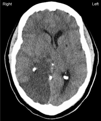
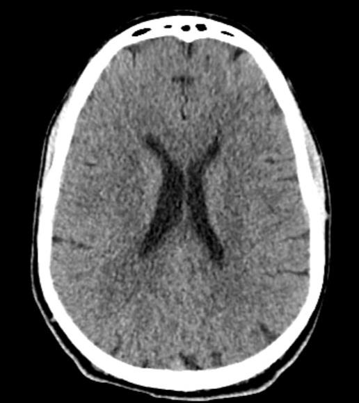
Compare the normal early NCCT (infarct not yet visible) with the hyperdense MCA sign of an acute large-vessel occlusion:
DWI-MRI is the most sensitive sequence, lighting up the infarct as a bright (restricted-diffusion) signal within minutes of onset:
Quantify severity using the full NIHSS scoring criteria, which grades 11 neurological domains to determine candidacy for reperfusion.
Differential Diagnosis
Several reversible or non-vascular conditions produce acute focal deficits. Always exclude hypoglycemia and hemorrhage first, and remember that a TIA produces identical deficits that resolve completely (classically <1 hour) without infarction on DWI.
| Stroke Mimics and Distinguishing Clues | |
|---|---|
| Mimic | Distinguishing clue |
| Hypoglycemia | Low fingerstick glucose; resolves with dextrose |
| Seizure / Todd paralysis | Postictal transient deficit; witnessed convulsion |
| Complicated migraine | Gradual, spreading aura; headache history |
| Bell palsy | Forehead involved (LMN); isolated facial weakness |
| Hypertensive encephalopathy | Global, non-focal symptoms; markedly elevated BP |
| Brain tumor / abscess | Subacute, progressive course; mass on imaging |
| Conversion disorder | Non-anatomic signs; normal imaging |
Management
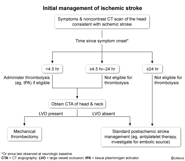
Acute reperfusion is organized around strict time windows. After airway/breathing/circulation are secured and NCCT excludes hemorrhage:
- Step 1 – IV thrombolysis: Alteplase (tPA) 0.9 mg/kg IV (max 90 mg), 10% as a bolus then the remainder over 60 minutes, within 4.5 hours of onset. Tenecteplase 0.25 mg/kg IV bolus (max 25 mg) is an accepted alternative. Confirm no exclusion criteria first.
- Step 2 – Blood pressure target: before tPA, lower BP to <185/110 mmHg (e.g., IV labetalol or nicardipine); maintain <180/105 for 24 h after. If the patient is not a lytic candidate, allow permissive hypertension and treat only if BP >220/120 mmHg.
- Step 3 – Mechanical thrombectomy: for proven large-vessel occlusion, perform endovascular thrombectomy up to 24 hours from onset in selected patients with favorable perfusion imaging.
- Step 4 – Antiplatelet: aspirin 325 mg within 24–48 h (delay until 24 h after tPA). For minor stroke/high-risk TIA, give short-term dual antiplatelet (aspirin + clopidogrel) for 21 days.
| Important – فكرة سؤال | |
A patient presenting beyond 4.5 hours is too late for IV thrombolysis but may still qualify for mechanical thrombectomy up to 24 hours if CT angiography confirms a large-vessel occlusion and perfusion imaging shows salvageable penumbra (DAWN / DEFUSE-3 criteria). Do not withhold vascular imaging just because the tPA window has closed. |
تذكر |
tPA eligibility is governed by rigid criteria; review the full tPA inclusion and exclusion criteria and the stroke blood-pressure management targets for the exact thresholds tested on exams.
The complete time-based decision pathway (tPA vs. thrombectomy) is summarised in this algorithm:
Secondary prevention (chronic):
- Antiplatelet (aspirin or clopidogrel) for non-cardioembolic stroke; high-intensity statin for all.
- Anticoagulation (DOAC or warfarin) for atrial fibrillation.
- Risk-factor control: BP, diabetes, smoking cessation.
- Carotid endarterectomy for symptomatic carotid stenosis 70–99%.
Complications & Prognosis
Complications
- Hemorrhagic transformation — most feared complication of reperfusion/thrombolysis.
- Cerebral edema & herniation — "malignant" MCA infarction peaking at 2–5 days.
- Seizures, post-stroke depression, and spasticity.
- Immobility-related: aspiration pneumonia, DVT/PE, pressure ulcers.
| Note – ملاحظة | |
Malignant MCA infarction peaks at 2–5 days as cytotoxic edema maxes out, causing herniation. In patients <60 years with large MCA infarcts and declining consciousness, decompressive hemicraniectomy is life-saving and reduces mortality. |
ملاحظة |
Prognosis
- The admission NIHSS is the strongest early predictor of outcome — higher scores correlate with worse disability and mortality.
- Earlier reperfusion = better outcome; every minute of delay costs viable neurons.
- Early multidisciplinary rehabilitation maximizes functional recovery; recurrence risk is highest in the first weeks, underscoring prompt secondary prevention.
Mnemonics
Consolidated high-yield memory hooks for acute ischemic stroke:
| Recognizing stroke — "BE-FAST" | |
|
جملة تذكرية |
| Modifiable stroke risk factors — "HEADS" | |
|
جملة تذكرية |
| Absolute tPA contraindications — "NO tPA" | |
|
جملة تذكرية |
Key Points for Exams – نقاط مهمة للامتحانات
- Ischemic stroke = ~85–87% of all strokes; hypertension is the most important modifiable risk factor and atrial fibrillation the classic cardioembolic source.
- First test = non-contrast head CT to exclude hemorrhage; DWI-MRI is the most sensitive for acute infarct. Always check a fingerstick glucose.
- IV alteplase 0.9 mg/kg (max 90 mg) within 4.5 hours; BP must be <185/110 before lysis.
- Mechanical thrombectomy up to 24 hours for large-vessel occlusion with salvageable penumbra.
- If not a lytic candidate → permissive hypertension; treat BP only if >220/120. Give aspirin 325 mg (delay 24 h after tPA).
- MCA → face/arm > leg + aphasia/neglect; ACA → leg > arm; PCA → homonymous hemianopia with macular sparing.
- Lacunar syndromes have no cortical signs (hypertension/diabetes, lipohyalinosis).
- Secondary prevention: antiplatelet + high-intensity statin; anticoagulate for AF; carotid endarterectomy for symptomatic 70–99% stenosis.
| Key Points for Exams | |
|
Note |
احصل على التجربة الكاملة
اشترك للوصول لفيديوهات الشرح التفصيلي والبطاقات التعليمية التفاعلية وأسئلة الممارسة مع تتبع التقدم.