SUMMARY
Gastrointestinal stromal tumors (GISTs) are the most common mesenchymal (sarcomatous) tumors of the gastrointestinal tract and account for the majority of GI sarcomas, with an incidence of 6–15 cases per million per year. They arise from cells phenotypically similar to the interstitial cells of Cajal (the gut pacemaker cells) and are driven by activating mutations in the KIT or PDGFRA receptor tyrosine kinases; c-Kit (CD117) is the defining immunohistochemical marker.
The stomach is the most common primary site and the liver is the most common site of metastasis. Once regarded as chemo-resistant, GISTs are now managed by complete surgical resection for localized disease and by the tyrosine-kinase inhibitor imatinib for locally advanced or metastatic disease, with sunitinib reserved for imatinib failure.
Pathogenesis & Molecular Biology
Cell of origin & immunohistochemistry
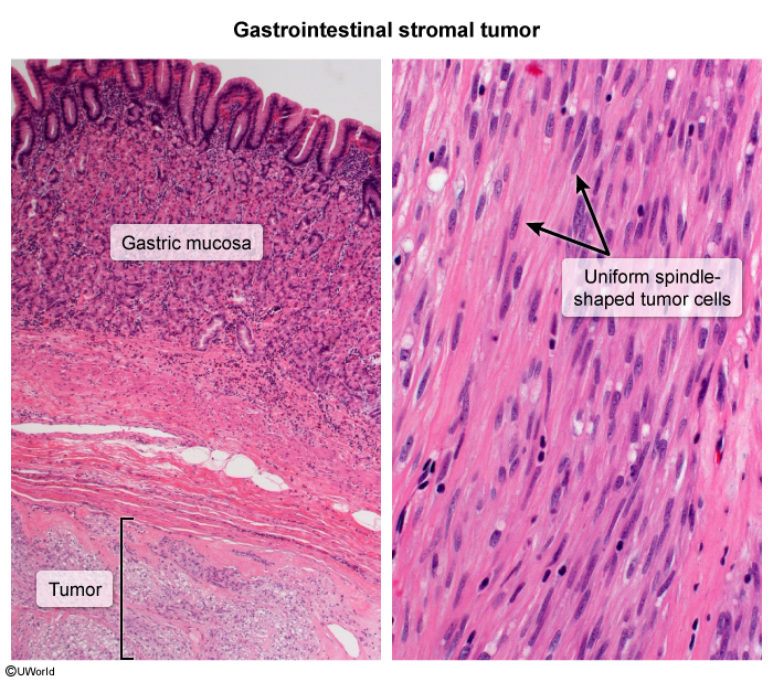
GISTs arise from cells phenotypically similar to the interstitial cells of Cajal, the intestinal pacemaker cells. Tumor cells characteristically express CD34 and c-Kit (CD117) — the latter being the defining immunohistochemical marker. Histologically, GISTs are composed of uniform spindle cells arranged in fascicles.
Driver mutations
GISTs are defined by activating (constitutive) mutations of the KIT or PDGFRA receptor tyrosine kinases, producing ligand-independent signalling. KIT mutations account for ~80% of cases and PDGFRA mutations for 5–10%; the remainder are wild-type. The key mutation sites are KIT exon 11, KIT exon 9, and PDGFRA exon 18. Mutation type is the single strongest predictor of response to imatinib, so the following master table is the reference point for GIST molecular biology.
| Molecular Biology of GIST — Mutations, Frequency & Imatinib Response | |
| KIT mutations | ~80% of GIST cases |
| KIT exon 11 | Most common KIT subtype. Best response to imatinib — 83.5%. Constitutive tyrosine-kinase activation. |
| KIT exon 9 | Less common KIT subtype. Lower response to imatinib — 47.8%. Frequent primary resistance. |
| PDGFRA mutations | 5–10% of GIST cases |
| PDGFRA exon 18 | Often resistant to imatinib. Constitutive activation. |
| Wild-type | Remaining cases |
| No detectable KIT/PDGFRA mutation | Less responsive to imatinib, but absence of mutation does NOT exclude GIST if morphology is typical. |
Important: The absence of a detectable mutation does NOT exclude GIST if the morphology is typical (wild-type GISTs). The predictive response figures above (83.5% for exon 11 vs 47.8% for exon 9) derive from phase 2 data and were confirmed in EORTC-62005.
Epidemiology & Clinical Presentation
Epidemiology & distribution
GISTs can arise anywhere along the GI tract but most commonly involve the stomach (60%) and small intestine (30%). Gastric GISTs carry a more favourable prognosis than those arising elsewhere.
Clinical presentation
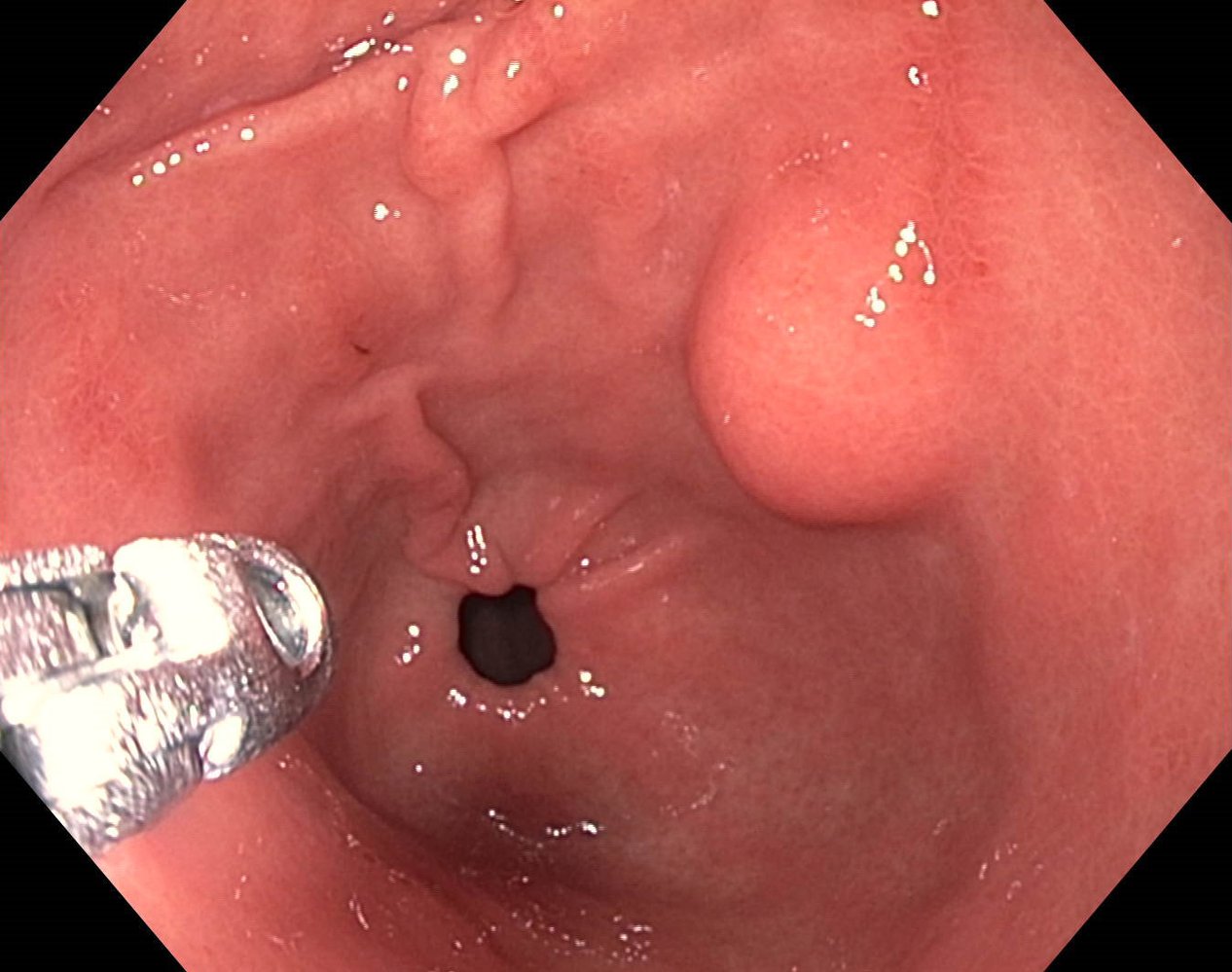
Many GISTs are incidental findings on upper endoscopy or CT. When symptomatic, patients present with early satiety, abdominal pain, or GI bleeding. Endoscopically a GIST typically appears as a submucosal mass with intact overlying mucosa.
Metastatic pattern
Metastasis is predominantly hematogenous to the liver (most common) and into the peritoneal/abdominal cavity; lymphatic spread is rare — the very fact that underlies the surgical principle of omitting lymphadenectomy.
| Clinical Features — Symptoms & Metastatic Pattern | |
| Detection | Frequently an incidental finding on upper endoscopy or CT |
| Symptoms | Early satiety; abdominal pain; GI bleeding |
| Common sites | Stomach (60%); small intestine (30%); may arise anywhere in the GI tract |
| Metastatic spread | Liver (most common); peritoneal/abdominal cavity; lymphatic spread is rare |
Radiologic Assessment
Imaging modality of choice
Contrast-enhanced CT of the abdomen and pelvis is the imaging modality of choice. It evaluates the primary tumor, peritoneal spread, and liver metastases. A primary gastric GIST typically appears as a large, exophytic, well-circumscribed mass that may show central necrosis.
FDG-PET
FDG-PET is useful for assessing baseline metabolic activity, detecting early metastases, and monitoring response to therapy — including early metabolic response to imatinib, which precedes any change in size on CT. A baseline PET is required before starting therapy if PET is to be used for response monitoring.
| ملاحظة – Note | |
|
A baseline PET scan must be performed before starting Imatinib if PET is to be used for monitoring metabolic response. |
ملاحظة |
Choi criteria
The Choi criteria are CT-based, evaluating tumor size and change in tumor density. They are used for assessing treatment response and prognosis prediction, capturing responses (density drop from tumor necrosis) that classic size-only criteria miss.
Management — Localized Disease
For localized GIST, complete surgical resection with negative margins (R0) is the treatment of choice. Because spread is hematogenous and not lymphatic, the operation is deliberately conservative — wide margins, extended anatomic resection, and lymphadenectomy are not required, and total gastrectomy is rarely necessary.
| Localized GIST — Management Algorithm | |
|---|---|
| Step 1 — Surgery | |
| Approach | Surgical principles |
| Complete R0 resection (negative margins) — treatment of choice | No wide margins, no extended anatomic resection, NO lymphadenectomy; total gastrectomy rarely required |
| Step 2 — Risk assessment | |
| Tool | Basis |
| NIH & AFIP risk-stratification systems | Tumor size, mitotic count, and site (AFIP) |
| Step 3 — Adjuvant therapy | |
| Candidate | Agent |
| High-risk resected tumors | Adjuvant imatinib |
Survival outcomes
- Overall 5-year survival: 20–44%
- 5-year survival after complete resection (early stage): up to 75%
- Disease-specific survival after complete resection: 54%
- Median survival in metastatic disease: ~20 months
Prognostic factors & risk stratification
Prognosis is driven chiefly by tumor size, mitotic rate, and location. These parameters feed the NIH and AFIP risk-stratification systems (AFIP additionally incorporates site), which are used to select candidates for adjuvant therapy.
| Prognostic Factors & Risk Stratification | |
| Tumor characteristics | Prognostic factor |
| Tumor size | Larger size → worse prognosis |
| Mitotic rate (per HPF) | Stratified as <5, 5–10, >10 — higher count → worse prognosis |
| Tumor location | Gastric → better prognosis than small-intestinal GIST |
| Risk stratification | System |
| NIH & AFIP | AFIP adds tumor site; based on size + mitotic count → select candidates for adjuvant therapy |
Management — Metastatic Disease & Targeted Therapy
First-line: imatinib
Imatinib (Gleevec, STI571) — a selective KIT tyrosine-kinase inhibitor — is first-line for locally advanced or metastatic GIST, dosed at 400 mg orally once daily. In the EORTC phase 1 study 53% achieved a partial response; a multicenter international trial of 147 patients (400 mg vs 600 mg daily) showed a 54% objective response with 14% progression and no dose–response difference.
Duration of therapy
Imatinib is continued as long as there is no progression. Stopping at 1 year worsens progression-free survival (~6 months vs 18 months); the optimal duration is not fully established.
Resistance
Primary resistance is progression within 6 months and is typical of KIT exon 9, PDGFRA exon 18, and wild-type tumors. Secondary resistance is progression after >6 months, usually due to acquired secondary mutations.
Escalation workflow
- Step 1: Start imatinib 400 mg orally once daily; continue while the tumor responds.
- Step 2: On progression/resistance → dose escalation (e.g., 400 → 600 mg daily).
- Step 3: If still progressing or intolerant → switch to sunitinib 50 mg orally once daily, 4 weeks on / 2 weeks off.
Second-line: sunitinib
Sunitinib is a multikinase inhibitor targeting VEGFR, PDGFRA, KIT, and FLT3, giving combined antiangiogenic and antiproliferative action. In a phase 3 trial the median time to progression was 27.3 weeks vs 6.4 weeks with placebo, and it is approved for imatinib-resistant or imatinib-intolerant GIST. The table below consolidates the two agents.
| Targeted Therapy Workflow — Imatinib vs Sunitinib | ||
|---|---|---|
| Feature | Imatinib (first-line) | Sunitinib (second-line) |
| Target | Selective KIT tyrosine-kinase inhibitor | Multikinase inhibitor: VEGFR, PDGFRA, KIT, FLT3 (antiangiogenic + antiproliferative) |
| Dose | 400 mg orally once daily (escalate on progression) | 50 mg orally once daily, 4 weeks on / 2 weeks off |
| Indication | First-line locally advanced or metastatic GIST | Imatinib-resistant or imatinib-intolerant GIST |
| Key trial data | 54% objective response (400 vs 600 mg — no dose–response difference) | Median time to progression 27.3 vs 6.4 weeks (placebo) |
| Top adverse effects | Diarrhea, nausea, periorbital edema, muscle cramps, fatigue; GI bleeding (5%, from rapid tumor necrosis) | Hand-foot syndrome, hypertension, cardiotoxicity, hypothyroidism (plus diarrhea/fatigue/nausea) |
Safety: Grade 3–4 toxicity occurs in ~21% of imatinib-treated patients; GI bleeding (~5%) can follow rapid tumor necrosis, so multidisciplinary follow-up including a surgeon is required.
Key Points for Exams
- GIST = CD117 (c-Kit)–positive mesenchymal tumor; arises from interstitial cells of Cajal (also CD34+).
- Most GISTs arise in the stomach (60%); small intestine is second (30%).
- Liver = most common site of metastasis; spread is hematogenous — lymphatic spread is rare.
- KIT exon 11 → best imatinib response; KIT exon 9 → lower; PDGFRA exon 18 / wild-type → often resistant.
- Localized disease: complete R0 resection; no lymphadenectomy and no wide margins required.
- Imatinib is first-line for metastatic disease and must be continued while effective.
- Sunitinib = second-line for imatinib-resistant/intolerant disease.
- FDG-PET detects early metabolic response to imatinib (obtain a baseline before therapy); Choi criteria add tumor density to size.
| Important – فكرة سؤال | |
|
A CD117 (c-Kit)–positive spindle-cell mesenchymal tumor of the GI tract = GIST. Imatinib response is predicted by mutation type: KIT exon 11 = best response; KIT exon 9 = lower; PDGFRA exon 18 and wild-type = often resistant. |
تذكر |
| GIST Memory Hooks – جملة تذكرية | |
|
جملة تذكرية |
احصل على التجربة الكاملة
اشترك للوصول لفيديوهات الشرح التفصيلي والبطاقات التعليمية التفاعلية وأسئلة الممارسة مع تتبع التقدم.