شرح المدرسين

Summary
Allergic (atopic) disorders are driven by an exaggerated IgE-mediated type I (immediate) hypersensitivity response to harmless environmental allergens. Mast-cell degranulation releases histamine, leukotrienes, and prostaglandins, producing the characteristic triad of vasodilation, smooth-muscle contraction, and increased vascular permeability.
This lesson covers the four major IgE-mediated (Type I) allergic disorders: Anaphylaxis, Allergic Rhinitis, Atopic Dermatitis (Eczema), and Urticaria (Hives).
Overview of Allergic Disorders
Allergic (atopic) disorders share one engine: an IgE-mediated type I hypersensitivity reaction. They cluster in the same patient and family and often appear in sequence — the atopic march: atopic dermatitis in infancy → allergic rhinitis → asthma. A positive personal or family history of atopy is a recurring clue across every condition in this lesson.
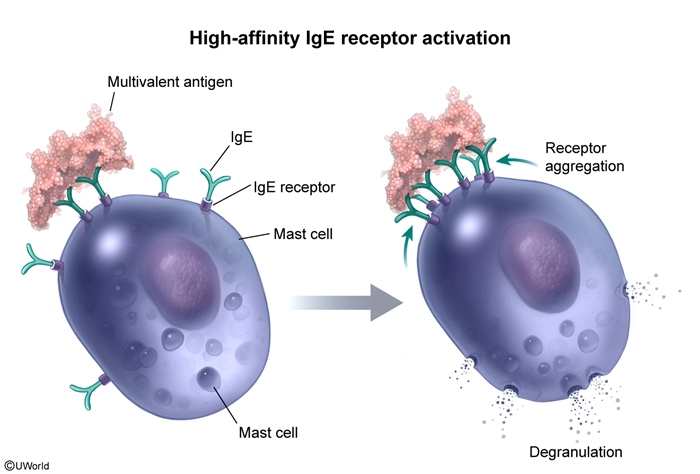
The shared mechanism is two-staged. During sensitization, allergen presentation drives a Th2 response (IL-4/IL-13) that switches B cells to produce allergen-specific IgE, which coats and primes mast cells and basophils. On re-exposure, allergen cross-links surface IgE and triggers degranulation, releasing histamine, leukotrienes, prostaglandins, and kinins. This produces an early phase (minutes — vasodilation, edema, wheal-and-flare, bronchospasm) and a late phase (hours — eosinophil-rich inflammation and induration).
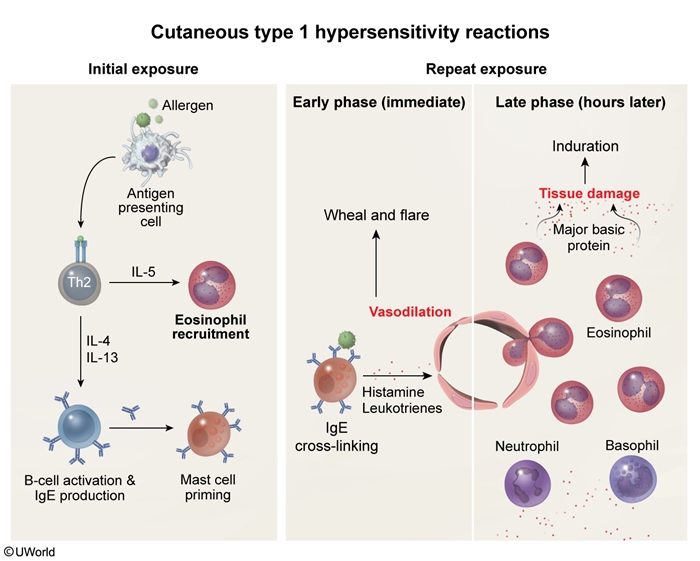
Type I is only one of four hypersensitivity mechanisms. See the Gell & Coombs hypersensitivity classification (Types I–IV) for how IgE-mediated allergy contrasts with the antibody-, immune-complex-, and T-cell–mediated types.
Anaphylaxis
Anaphylaxis is a potentially life-threatening, acute systemic IgE-mediated (type I hypersensitivity) reaction. It occurs most commonly to drugs, insect venom, foods, latex, and biologic agents. Antigen binding to IgE on the surface of primed mast cells and basophils releases potent mediators that affect vascular tone and bronchial reactivity.
Clinical presentation
Anaphylaxis involves two or more organ systems:
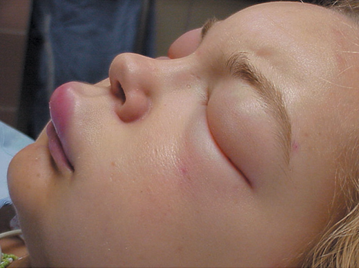
- Mucocutaneous — pruritus, flushing, urticaria, and angioedema (present in ~90% of cases).
- Respiratory — dyspnea and wheezing.
- Gastrointestinal — nausea, vomiting, diarrhea, and crampy abdominal pain.
- Cardiovascular — ranging from mild hypotension to shock.
| Mnemonic – Anaphylaxis: the 4 systems | |
|
‘Skin, Sats, Stomach, Systolic’ — the four systems anaphylaxis attacks:
Involvement of ≥2 systems after an exposure = anaphylaxis. |
جملة تذكرية |
Diagnosis
Diagnosis is clinical: characteristic signs and symptoms occurring within 30–90 minutes after exposure to the offending agent.
Management
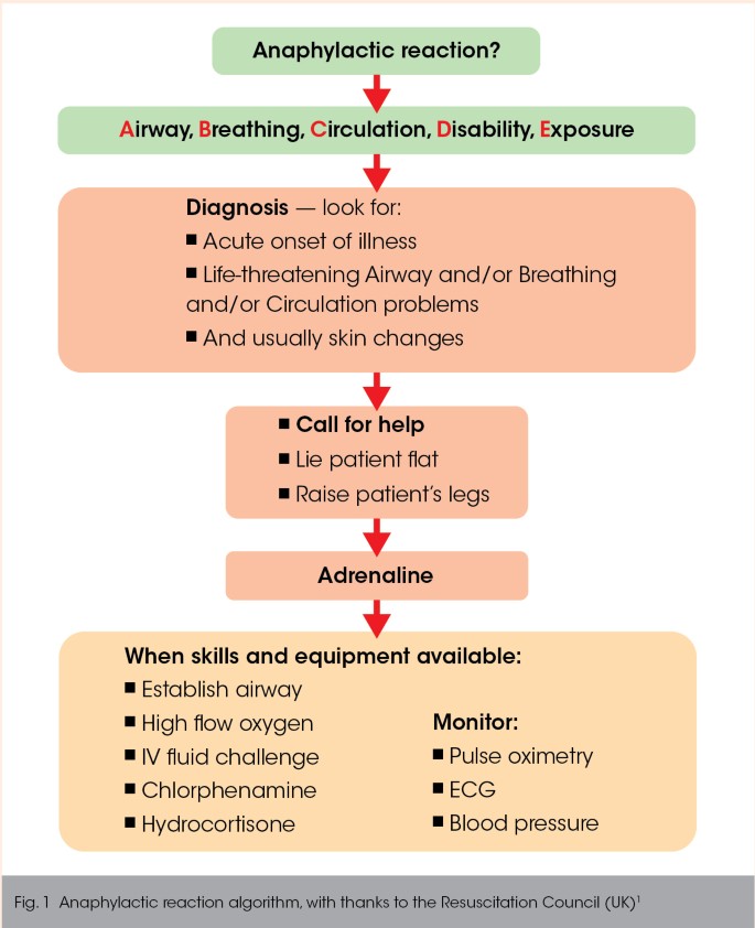
- Epinephrine is the principal treatment for the acute respiratory and cardiovascular complications — given IM into the mid-outer thigh.
- Systemic antihistamines, corticosteroids, and β-adrenergic agonists are used only as adjuncts to treat residual signs and symptoms. Refer to the anaphylaxis treatment algorithm for the immediate and adjunctive steps.
| Note | |
|
Exam trap: Always remember that the first-line treatment in anaphylaxis is IM Epinephrine (in the mid-outer thigh), not antihistamines or steroids. Antihistamines and steroids are only adjuncts and do not stop airway edema or shock. |
ملاحظة |
| Important – Question Idea | |
|
Urticaria alone ≠ anaphylaxis. Isolated hives/pruritus with stable vitals and no other system involved (e.g., a mild amoxicillin reaction) is treated with an H1 antihistamine — epinephrine and steroids are not required. The moment a second organ system is involved (respiratory, cardiovascular, or GI), it becomes anaphylaxis and demands IM epinephrine. |
تذكر |
Allergic Rhinitis
Allergic rhinitis is an IgE-mediated inflammatory response of the nasal mucosa to inhaled antigens. It is one of the most common allergic conditions of childhood, affecting 10–20% of children, and is commonly associated with asthma, chronic sinusitis, otitis media with effusion, and nasal polyps. Two subtypes are recognized:
| Seasonal vs Perennial Allergic Rhinitis | ||
|---|---|---|
| Feature | Seasonal | Perennial |
| Timing | Specific seasons | Across seasons (year-round) |
| Typical allergens | Tree, grass, and weed pollens (outdoor) | Dust mites, molds, animal dander (indoor) |
Pathophysiology
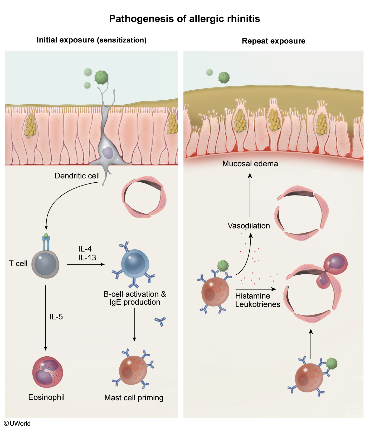
- Sensitization to airborne allergens induces IgE formation.
- Allergen-specific IgE binds mast cells and basophils in the nasal mucosa.
- On subsequent exposure (“priming”), primed mast cells degranulate within minutes and release histamine, leukotrienes, kinins, and prostaglandins, producing the IgE-mediated inflammatory response.
Clinical features
Sneezing, nasal congestion, rhinorrhea, nasal itching, and pale nasal mucosa. Classic exam signs include:
- Allergic shiners — dark circles under the eyes from venous congestion.
- Dennie-Morgan folds — creases under the eyes from chronic edema.
- Allergic salute — a transverse crease across the nasal bridge (between the upper two-thirds and lower one-third), from repeatedly pushing the nose up with the palm to relieve itching.
Diagnosis
Largely clinical. History may include recurrent otitis media, sinusitis, atopic dermatitis, and food or drug allergies. Serum: total IgE may be elevated. Special tests: allergen skin testing (prick or intradermal). The allergic rhinitis clinical signs and first-line treatment summary lays out the hallmark examination findings and management side by side.
Management
Allergen avoidance is the first step. Pharmacotherapy:
- Intranasal corticosteroids — most effective class for controlling symptoms (side effect: local irritation).
- Antihistamines — first-generation (e.g., diphenhydramine) are first-line but sedating and can impair academic performance; second-generation (cetirizine, fexofenadine, loratadine) are better tolerated but no more effective.
- Decongestants (e.g., pseudoephedrine) — relieve congestion via vasoconstriction; limit to <48–72 hours to avoid rebound rhinitis.
- Intranasal cromolyn, leukotriene receptor antagonists, and immunotherapy are additional options.
| Clinical Note | |
|
Intranasal corticosteroids are the most effective class for controlling symptoms. Decongestants (pseudoephedrine) should be used only for <48–72 hours to avoid rebound rhinitis (rhinitis medicamentosa). |
Note |
Atopic Dermatitis (Eczema)
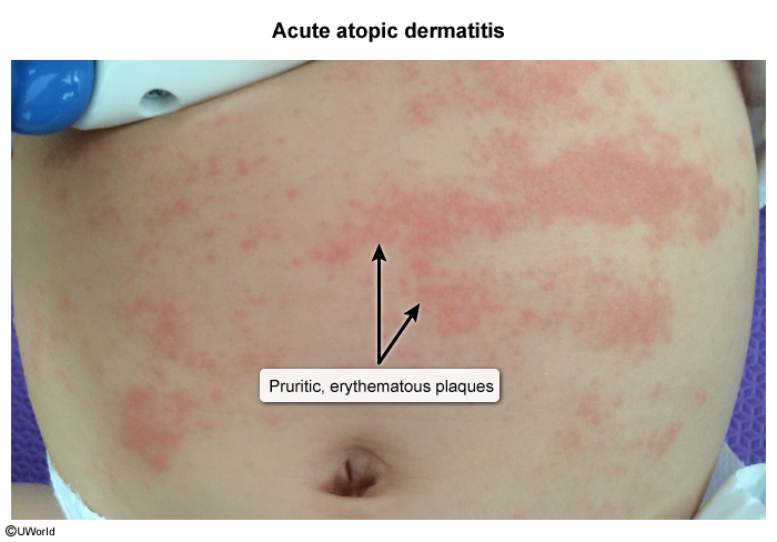
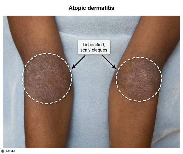
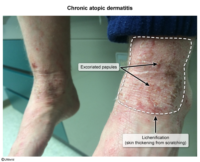
Atopic dermatitis (eczema) is a chronic inflammatory dermatitis characterized by dry skin and lichenification (skin thickening). The skin is overly sensitive to many stimuli that provoke pruritus, and the resulting scratching produces most of the visible skin changes. It affects 5–8% of children.
Clinical features
Onset is typically in early infancy, with 85% of patients showing signs before 5 years of age. It is often worse in winter or with temperature extremes, and there is usually a positive family history of atopy (eczema, asthma, or other allergic disease). Pruritus is universal.
| Mnemonic – Atopic Dermatitis | |
|
‘The itch that rashes’ — pruritus is universal and comes first; scratching drives the itch–scratch cycle that produces erythema, weeping, and eventually lichenification. The atopic triad travels together: Eczema + Asthma + Allergic rhinitis. |
جملة تذكرية |
| Atopic Dermatitis: Acute vs Chronic Skin Changes | ||
|---|---|---|
| Feature | Acute changes | Chronic changes |
| Primary skin findings | Erythema; weeping and crusting | Lichenification (from chronic itching); dry, scaly skin |
| Secondary changes | Secondary infection — bacterial (S. aureus), viral (HSV) | Pigmentary changes — usually hyperpigmentation, less often hypopigmentation |
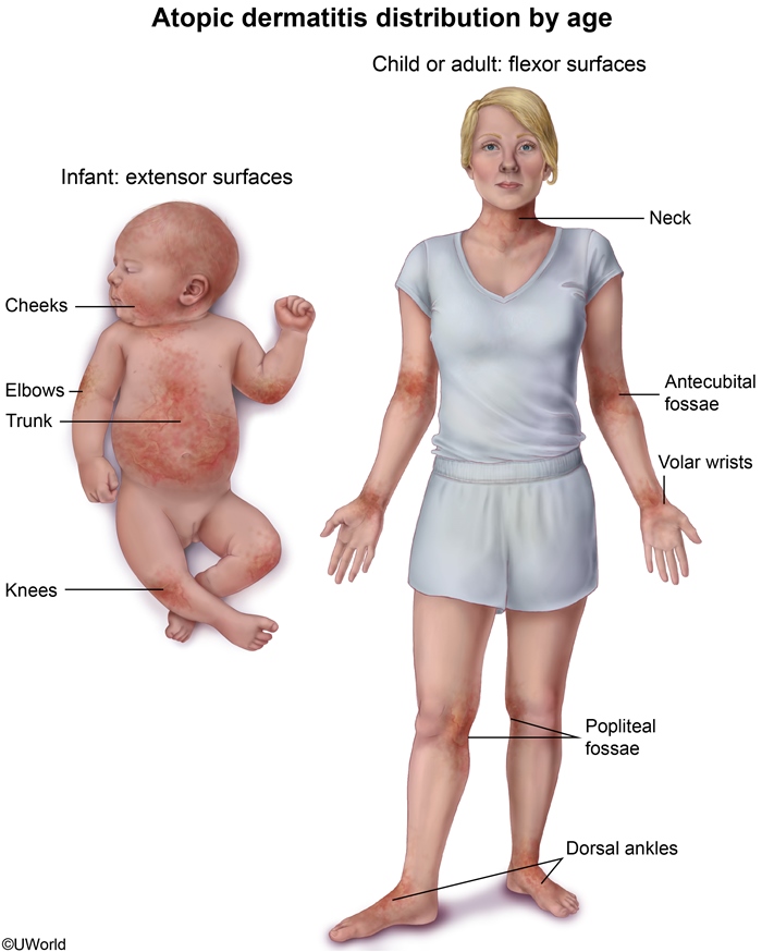
Distribution by age
| Atopic Dermatitis: Distribution by Age | |
|---|---|
| Infantile | |
| Predominant sites | Hallmark |
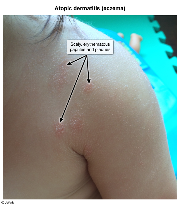
| Face, scalp, and trunk; extensor surfaces > flexural surfaces | Acute weeping erythema |
| Early childhood | |
| Predominant sites | Hallmark |
| Flexural surfaces (antecubital and popliteal fossae) | Lichenification |
| Late childhood | |
| Predominant sites | Hallmark |
| More localized patches | Tendency toward remission |
| Exam Trap – Eczema distribution | |
|
In infants, eczema appears on the face, scalp, and extensor surfaces. However, as the child grows (early childhood), it shifts to the flexural surfaces (antecubital and popliteal fossae) with lichenification. Examiners frequently test on this age-related distribution shift. |
Note |
Diagnosis
Diagnosis is clinical: 3 of 4 major criteria are required, with minor criteria providing support.
| Diagnostic Criteria for Atopic Dermatitis | |
|---|---|
| Feature Category | Clinical Features |
| Major features |
— In children under 2 years: face, trunk, and extensor surfaces
|
| Minor features |
|
Management
Management: avoid known triggers; use low- to medium-potency topical corticosteroids (systemic corticosteroids only in severe cases); apply antihistamines at bedtime to break the itch–scratch cycle; take tepid-water baths, then blot dry and apply skin lubricants/emollients to restore the barrier.
Urticaria (Hives)
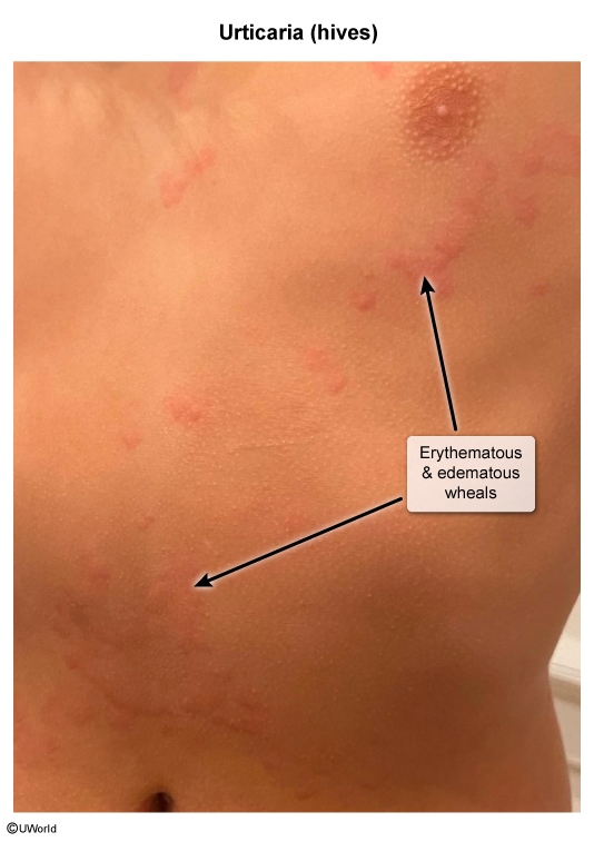
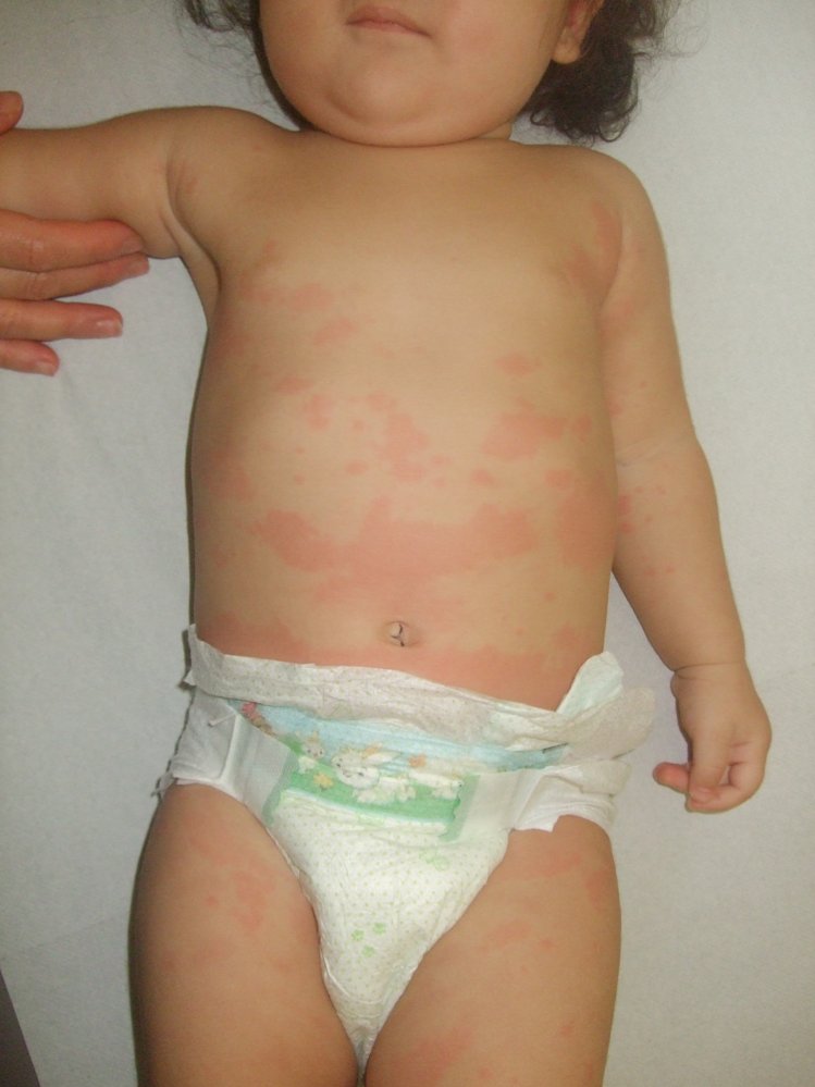
Urticaria (hives) consists of circumscribed, raised, evanescent (transient) areas of edema and erythema that are almost always pruritic.
Lesions are usually symmetric and migratory, with individual wheals typically resolving within 24 hours. It reflects mast cell degranulation with histamine release causing superficial dermal edema.
Etiology
| Causes of Urticaria: Acute vs Chronic (>6 weeks) | |
| Acute urticaria | Common triggers |
| Idiopathic | No identifiable cause (most common) |
| Drugs | Penicillin, aspirin, nonsteroidal anti-inflammatory drugs (NSAIDs) |
| Foods | Eggs, shellfish, milk, nuts |
| Contactants | Animal dander, latex |
| Infections | Group A β-hemolytic streptococcal pharyngitis, infectious mononucleosis, Mycoplasma pneumoniae, hepatitis, coxsackievirus |
| Insect venoms | Bee, wasp, hornet stings |
| Transfusion reactions | Blood product administration |
| Physical triggers | Heat, cold, skin pressure, exercise |
| Chronic urticaria (>6 weeks) | Underlying associations |
| Idiopathic | Most cases |
| Thyroid disease | Autoimmune thyroiditis |
| Malignancy | Lymphoma, solid tumors |
| Rheumatologic disease | SLE, rheumatoid arthritis |
Management
Avoid precipitating factors. In chronic urticaria, identify any underlying systemic disease. Antihistamines are the mainstay of therapy.
| Clinical Note – Urticaria | |
|
Clinical Note: Chronic urticaria refers to wheals lasting >6 weeks, with most cases being idiopathic. |
Note |
Key Points for Exams
All four entities are type I (IgE-mediated) hypersensitivity reactions sitting along the atopic march (eczema → allergic rhinitis → asthma). The exam-relevant discriminators are the threshold for anaphylaxis, the signs of allergic rhinitis, the age shift in eczema distribution, and the mainstay for urticaria.
| Important – Question Idea | |
|
تذكر |
Consolidated mnemonics
| Anaphylaxis — the 4 systems | |
| S = Skin (mucocutaneous) S = Sats (respiratory) S = Stomach (GI) S = Systolic (cardiovascular) ≥2 systems = anaphylaxis |
جملة تذكرية |
| Atopic dermatitis & Atopic triad | |
| The itch that rashes — pruritus first, then the itch–scratch cycle E = Eczema A = Asthma A = Allergic rhinitis |
جملة تذكرية |
احصل على التجربة الكاملة
اشترك للوصول لفيديوهات الشرح التفصيلي والبطاقات التعليمية التفاعلية وأسئلة الممارسة مع تتبع التقدم.